Figure 4.
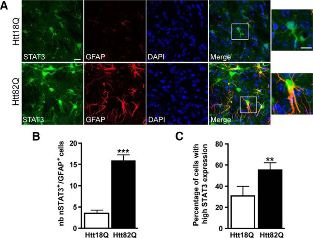
The JAK/STAT3 pathway is activated in reactive astrocytes in the mouse model of HD. A, Images of brain sections showing double staining for GFAP (red) and STAT3 (green) on mouse brain sections, 6 weeks after the infection of striatal neurons with lenti-Htt18Q or lenti-Htt82Q. Astrocytes in the Htt82Q striatum are hypertrophic and express higher levels of STAT3 in their nucleus relative to resting astrocytes in the Htt18Q striatum. B, C, The number of nSTAT3+/GFAP+ cells (B) and the percentage of cells displaying strong staining for STAT3 (C) are significantly higher in the Htt82Q striatum than in the Htt18Q striatum. n = 6. **p < 0.01, ***p < 0.001. Scale bars: 20 μm; enlargements, 5 μm.

