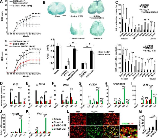
Figure 1.
Therapeutic benefits of SHEDs or SHED-CM for SCI. A, Hindlimb functional recovery after spinal cord contusion. Top, SHED transplantation, n = 7; PBS, n = 5. Bottom, SHED-CM, n = 15; BMSC-CM, n = 7; serum-free DMEM, n = 10. ANOVA with Tukey's post hoc test. B, Sudan black B staining of axial spinal cord sections 3 mm caudal to the epicenter 8 weeks after SCI and quantification of gray and white matter areas 3 mm caudal to the epicenter. ANOVA with Tukey's post hoc test (n = 3 rats per group). C, Quantification of the 5-HT-positive nerve fibers in sagittal sections of the spinal cord, 8 weeks after SCI. x-Axis indicates specific locations along the spinal cord rostrocaudal axis. Results are expressed relative to the value in sham-operated rats at the Th9 level. ANOVA with Tukey's post hoc test (n = 3 rats per group). D–K, qPCR analysis of the indicated mRNAs in CM-treated spinal cords over time. Results are expressed relative to the level in the sham-operated model. ANOVA with Tukey's post hoc test (±2 mm from epicenter, n = 3 rats per group). L, Representative images of immunohistological staining of microglia/macrophages surrounding the lesion (±1 mm from epicenter) 72 h after SCI. Antibodies used and treatment groups are indicated at top and left, respectively. CD206+ cells in SHED-CM-treated spinal cord highly coexpressed IL-10. Arrowheads indicate CD206+/Iba1+ cells. M, Quantification of CD206+/Iba1+ cells in treated spinal cords. ANOVA with Tukey's post hoc test (n = 3 rats per group and 5 sections per animal). Scale bars: B, 500 μm; L, 100 μm. Mean ± SD (A–C and M) and mean ± SEM (D–K). *p < 0.05, **p < 0.01, ***p < 0.001.

