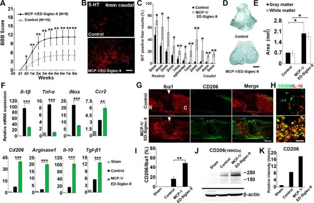
Figure 7.
MCP-1/ED-Siglec-9 activates an anti-inflammatory M2 response and promotes functional recovery after SCI. A, Recovery of hindlimb locomotion after SCI. PBS, n = 10; MCP-1/ED-Siglec-9, n = 9. ANOVA with Tukey's post hoc test. B, Immunohistological images of 5-HT-positive nerve fibers in sagittal sections of the injured spinal cord 4 mm caudal from the epicenter, 8 weeks after SCI. C, Quantification of 5-HT-positive nerve fibers from 5 mm rostral to 5 mm caudal. Unpaired two-tailed Student's t test (n = 3 rats per group). D, E, Sudan black B staining of axial spinal cord sections 3 mm caudal from the epicenter 8 weeks after SCI, and quantification of gray and white matter areas 3 mm caudal from epicenter. ANOVA with Tukey's post hoc test (n = 3 rats per group). F, Levels of the indicated mRNAs in the spinal cord 72 h after SCI. ANOVA with Tukey's post hoc test (n = 3 rats per group). G, H, Immunohistological images of sagittal spinal cord sections 72 h after SCI. Left, Treatment; top, antibodies used. I, Quantification of CD206+/Iba1+ cells in the spinal cord. An unpaired two-tailed Student's t test (n = 3 rats per group and 5 sections per animal). J, Immunoblot analysis for CD206 protein 72 h after SCI. K, Expression intensity analysis of CD206 protein bands. Scale bars: D, G, 500 μm; B, H, 100 μm. Mean ± SD (A, C, E, I) and mean ± SEM (F). *p < 0.05; **p < 0.01; ***p < 0.001.

