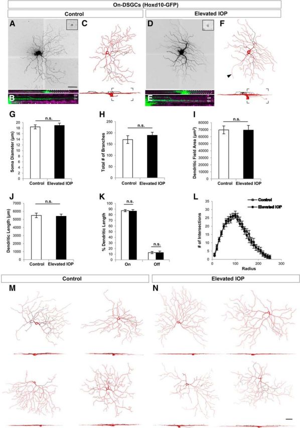
Figure 5.

On DSGCs are structurally resistant to the early phase of elevated IOP. A–F, Same format as for Figure 3 but GFP cells are On DSGCs from Hoxd10-GFP mice. G–K, Quantification of various morphological parameters examined for RGCs in control (white bars) and IOP-elevated (black bars) eyes. No significant differences (n.s.) were found for any of the morphological parameters studied, including soma diameter (G), total number of branches (H), dendritic field area (I), dendritic length (J), as well as the percentage dendritic length in the On versus Off sublaminae (K). L, Moreover, no changes were noted when examining the overall dendritic architecture using Sholl ring analysis. For G–L, error bars represent ±SEM. M, N, Representative examples of On DSGCs in control (M) and IOP-elevated retinas (N). For C, F, M, and N, red represents the soma, axon (arrowhead), and dendrites found in the On sublamina, while dendrites present in the Off sublamina are depicted in black. Scale bars: A, N, 50 μm.
