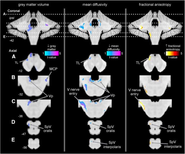
Figure 3.
Regions of structural difference in subjects with PTN compared with pain-free control subjects overlaid onto template axial and coronal T1-weighted images. Slice locations in MNI space are indicated at the lower left of each image. Left, Regional gray matter volume decreases (cool color scale) in subjects with PTN compared with pain-free controls. Subjects with PTN have lower gray matter volumes in the ipsilateral (to highest ongoing pain) SpV oralis, ipsilateral Vp, contralateral MCP, and the ipsilateral TL. Middle, Regional MD decreases (cool color scale) in subjects with PTN compared with pain-free controls. Subjects with PTN have lower MD in the ipsilateral SpV oralis and interpolaris, ipsilateral V nerve entry, bilateral Vp, and ipsilateral TL. Right, Regional FA increases (hot color scale) in subjects with PTN compared with pain-free controls. Subjects with PTN have higher FA in the ipsilateral SpV oralis and interpolaris, ipsilateral V nerve entry, and the ipsilateral TL.

