


Figure 2.
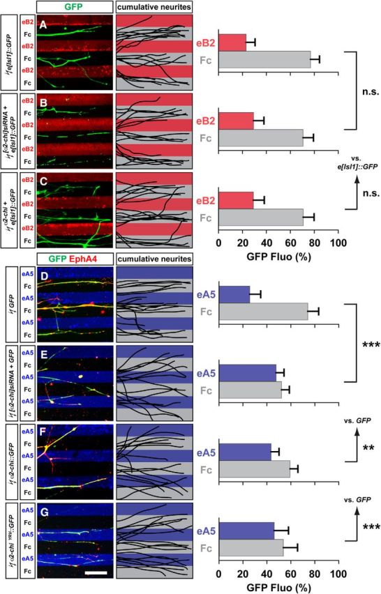
α2-chimaerin function is required for EphA forward signaling in cultured LMC neurons. A–C, Detection of medial (GFP+) LMC neurites of e[Isl1]::GFP (A), [α2-chi]siRNA + e[Isl1]::GFP (B), or α2-chi + e[Isl1]::GFP (C) on ephrinB2-Fc/Fc stripes and superimposed images of five representative explants from each experimental group highlighting the distribution of LMC neurites. Quantification of medial (GFP+) LMC neurites on first (red) and second (pale) stripes expressed as a percentage of total GFP signals. Minimal number of neurites and explants: 79 and 11 for all groups. D–G, Detection of lateral (EphA4+) LMC neurites of GFP (D), [α2-chi]siRNA + GFP (E), α2-chi::GFP (F), or α2-chiΔYRV::GFP (G) on ephrinA5-Fc/Fc stripes and superimposed images of five representative explants from each experimental group highlighting the distribution of LMC neurites. Quantification of electroporated lateral (EphA4+) LMC neurites on first (blue) and second (pale) stripes expressed as a percentage of total EphA4 signals. Minimal number of neurites and explants: 72 and 10 for all groups. eB2, ephrin-B2; eA5, ephrin-A5. Error bars indicate SD. ***p < 0.001; **p < 0.01; n.s., not significant; statistical significance computed using Student's unpaired t test; all values are mean ± SD. Scale bars in A–G, 50 μm.