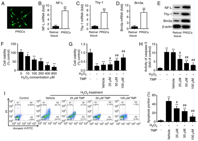
Figure 1.
TMP promotes cell viability and suppresses cell apoptosis in H2O2-treated PRGCs. (A) NF-L expression was detected by immunofluorescence (magnification, ×200). (B-D) The mRNA expression levels of NF-L, Thy-1 and Brn3a were measured by reverse transcription-quantitative PCR. (E) Protein expression of NF-L, Thy-1 and Brn3a as determined by western blotting. β-actin was used as a loading control. (F) PRGCs were treated with 10, 100, 200, 400 and 800 µM of H2O2 for 24 h, and then cell viability was determined by a CCK-8 assay. PRGCs were pre-treated with 25, 50 and 100 µM TMP for 24 h prior to exposure with 400 µM of H2O2. (G) Cell viability was determined by a CCK-8 assay. (H) The activity of caspase-3 was determined using a caspase-3 assay kit. (I) Apoptosis was detected via flow cytometric analysis. Data are presented as mean of three replicates ± standard deviation. *P<0.05, **P<0.01 vs. Control group, #P<0.05, ##P<0.01 vs. Vehicle + H2O2 group. CCK-8, Cell Counting Kit-8; FITC, fluorescein isothiocyanate; NF-L, neurofilament-L; PI, propidium iodide; PRGCs, primary retinal ganglion cells; Thy-1, thymus cell antigen 1; TMP, tetramethylpyrazine.

