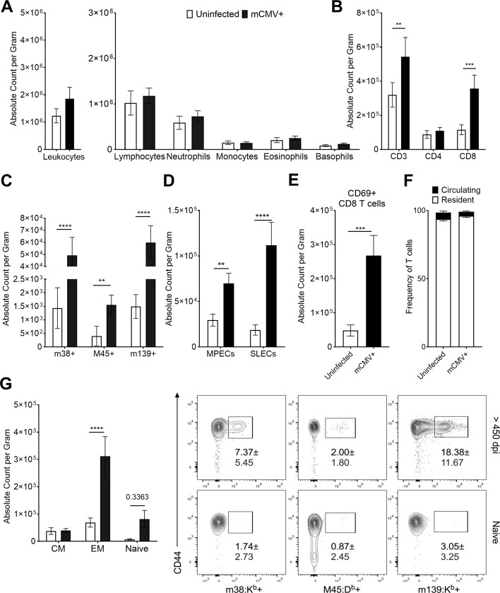
Fig 4. mCMV-specific T cells are maintained in adipose tissue for the lifespan of infection.
12-week-old C57BL/6J mice were i.p. injected with 105 pfu of mCMV and sacrificed >450d p.i. Stromal vascular fraction was analyzed by Drew Scientific HemaVet 950 and flow cytometry. (A) Total leukocytes were quantified by hemocytometer. (B) Flow cytometry analysis was used to quantify absolute numbers per gram of adipose tissue of CD3 T cells and gated on CD4 or CD8. (C) CD8 T cells were phenotyped based on expression of CD62L and CD44 and quantified. (D) CD44+ CD8 T cells were analyzed for expression of KLRG1 and CD127 to quantify number of MPECs and SLECs. (E) Total CD8 T cells were analyzed for surface expression of CD69. (F) Lifelong infected animals were injected i.v. with 3 μg of CD45 antibody to determine tissue residency of T cells. Frequency of in vivo and ex vivo stained animals is shown. (G) CD44+ CD8 T cells were analyzed for mCMV specificity by tetramer staining. (A-E and G) Data are pooled results of two independent experiments. n = 20 uninfected animals and 19 infected animals total. (F) Data are pooled results of two independent experiments with an n = 9 uninfected animals and n = 9 infected animals total. Frequencies shown in the dot plots represent SD. Error bars represent mean ± SEM. *p < 0.05; **p < 0.01; ***p < 0.001; **** p ≤ 0.0001 by unpaired two-tailed Mann-Whitney U test.

