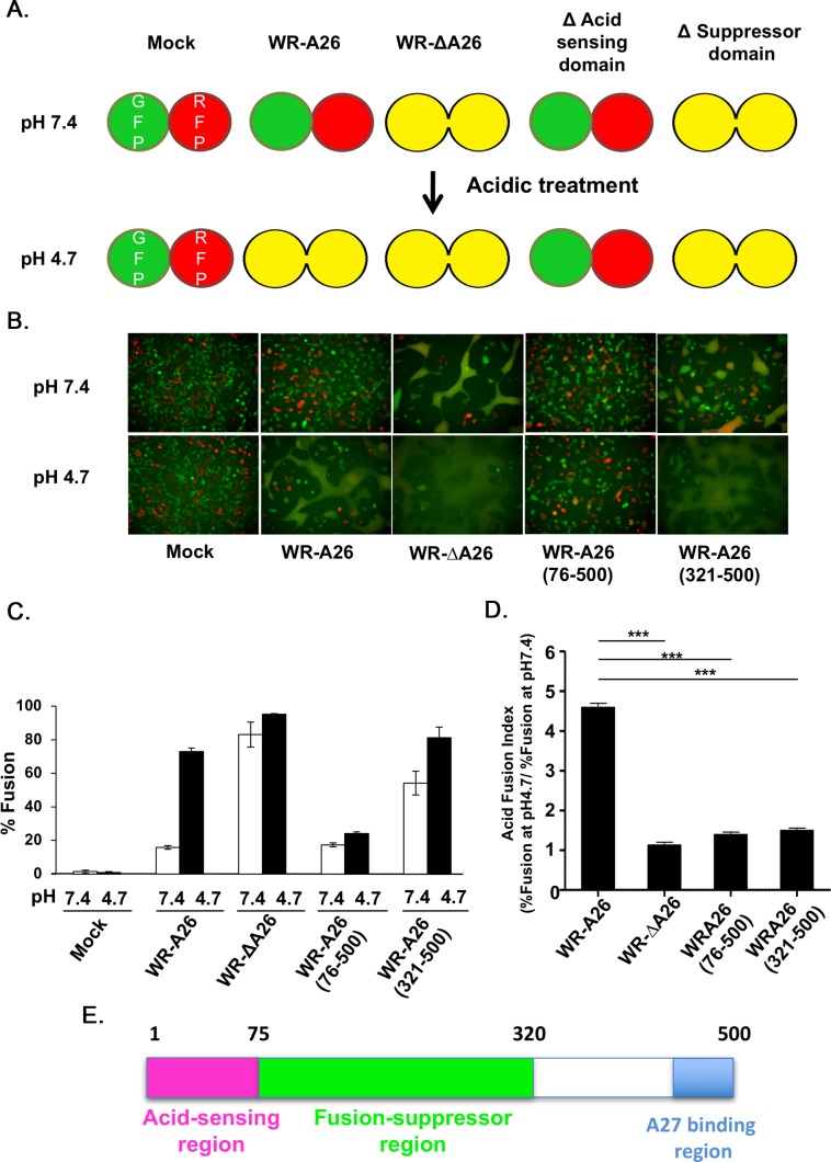
Fig 2. N-terminal region of A26 protein (aa 1–75) is important for endocytic-mediated MV membrane fusion at low pH.
(A). Schematic presentation of vaccinia MV-dependent cell-cell fusion at neutral and acidic pH. L cells expressing GFP and RFP were mixed at a 1:1 ratio during seeding. No cell fusion occurs at either pH in the absence of infection (Mock). When cells were infected with the endocytic WR-A26 virus, cell-cell fusion only occurred at low pH (which mimics endosomal environments) and no cell-cell fusion occurred at neutral pH (which allows plasma membrane fusion). In contrast, WR-ΔA26 virus does not require acidic environments to activate membrane fusion, so cell-cell fusion occurs at both acidic and neutral pH. Truncation of A26 protein to remove the acid-sensing/acid-sensitive domain (WR-A26(76–500)) renders the protein a constitutive fusion suppressor, so no fusion occurred at either pH. Further truncation of A26 protein to remove the fusion suppressor domain (WR-A26(321–500)) resulted in a virus phenotype like that of WR-ΔA26 in that it fuses at both pH. (B). Images of cell-cell fusion induced by various vaccinia MV infections. Single fluorescent cells described in (A) were infected with each virus at an MOI of 50 PFU per cell, washed with neutral or acidic pH buffer for 3 min, and then subjected to live-imaging to monitor cell-cell fusion at 37°C for 2 h. (C). Quantification of % cell fusion induced by each virus at neutral or low pH conditions as described in (B). Five images for each virus were recorded and the % fusion was calculated using the image area of GFP+RFP+ double-fluorescent cells divided by that of single-fluorescent cells. The experiments were repeated three times for each virus and bars represent standard deviation. (D). An “Acid Fusion Index” was calculated to represent the acid-dependence of each A26 deletion protein, i.e., the occurrence of A26 protein conformational change. The index for each virus was obtained by dividing the % of cell fusion at low pH (black bar in C) by that recorded for neutral pH (white bar in C). Endocytic WR-A26 virus demonstrated an Acid-Fusion Index of ~4.6, whereas the value for all other viruses was ~1, meaning these latter exhibit limited dependence on acidic environments for cell fusion. The experiments were repeated three times for each virus and the Student t-test was used for statistical analyses. *** p <0.001. (E). Schematic representation of A26 protein functional regions. The N-terminal region of A26 protein (aa 1–75, in pink) is important for acid sensitivity, whereas the middle region (aa 76–320, in green) is required for the fusion suppressor function. The C-terminal region (in blue) was previously shown to mediate binding to viral A27 protein during MV assembly [34, 35].

