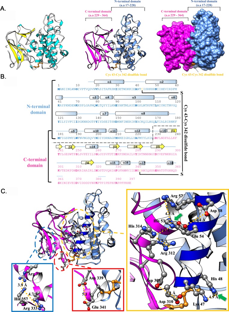
Fig 5. Crystal structure of recombinant A26 protein (aa 1–397).
(A). A ribbon diagram of the A261-397 structure. The α-helices and β-strands are colored in cyan and yellow, respectively. Two domains, NTD (aa 17–228) and CTD (aa229-364), are shown and highlighted in cornflower blue and magenta, respectively. (B). The secondary structural elements are shown above the amino acid sequences, with cyan cylinders and yellow arrows representing α-helices and β-strands, respectively. (C). The His-cation and AniAni pairs in the recombinant A261-397 structure. The amino acids that involve in His-cation and AniAni pairs are shown as ball and stick models. Green arrows show the position of His48 and His53.

