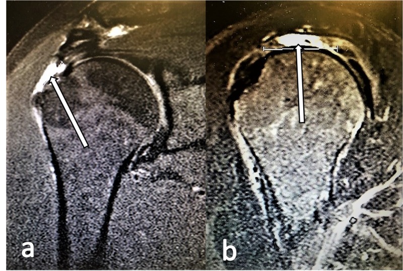
Figure 2. Supraspinatus tears depicted in MRI scans.
a. T2-weighted oblique coronal MRI view with a high-intensity signal (white arrow) revealing a full-thickness tear of the supraspinatus.
b. T2-weighted oblique sagittal MRI view with a high-intensity signal (white arrow) revealing a full-thickness tear of the supraspinatus.

