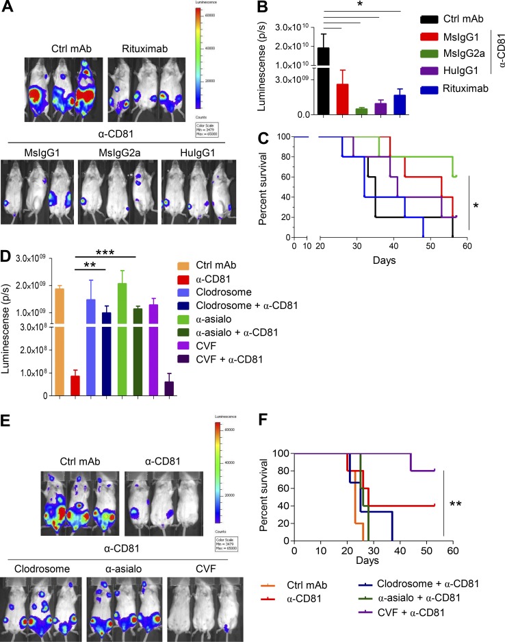
Figure 3.
5A6 eliminates Raji cells in vivo in a macrophage- and NK cell–dependent manner. (A–C) 1.5 × 10⁶ Raji-luciferase cells were injected i.v. into SCID-Beige mice. Mice were treated on day 5 after tumor challenge i.p. with 100 µg and weekly thereafter (four doses) with the indicated 5A6-based mAbs (MsIgG1, MsIgG2a, and chimeric HuIgG1); rituximab served as a positive control, and a nonspecific MsIgG1 served as a negative control. Ctrl, control. (A) Representative in vivo bioluminescence imaging (IVIS) on day 22 after challenge. (B) Bioluminescence quantification (Living Image) on day 29 (n = 5 mice in each group). (C) Survival was quantified when mice exhibited hindleg paralysis. (D–F) Macrophages from SCID-Beige mice were depleted with 200 µl of clodrosome liposomes (5 mg/ml) i.p. 4 h after tumor challenge and on day 5. NK cells were depleted by i.p. injection of 50 µg of anti-asialo GM1 on days −1 and 0 of tumor challenge and every 5 d thereafter for 3 wk. Complement was depleted by i.p. injection of 25 U CVF on the day of tumor challenge and on days 3, 6, and 9 after challenge. Mice (five per group) were injected with Raji-luc cells as in Figs. 1 A and 3 A and then treated with 100 µg of 5A6 MsIgG2a or isotype control on day 5 and every week thereafter for a total of four doses. (D) Bioluminescence quantification (Living Image) on day 20 after tumor challenge. (E) Representative in vivo bioluminescence imaging (IVIS) on day 20 after lymphoma injection. (F) Survival was quantified as in C. Error bars represent mean ± SEM. *, P < 0.0149; **, P < 0.0036; ***, P < 0.0001; Student’s t test (B and D; n = 5 mice in each group). *, P < 0.0154; **, P < 0.0022; log-rank (Mantel–Cox) test (C and F).

