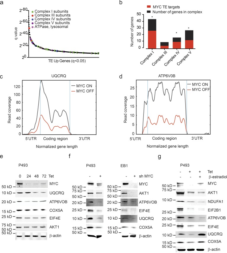
Figure 2.
MYC stimulates the translation of ETC proteins. (a) MYC-dependent mRNAs (TE up; ranked by significance) reveal a preponderance of ETC components: complex I genes (green), complex III genes (red), complex IV genes (blue), complex V genes (purple), and lysosomal ATPase (pink); n = 3 biological replicates in each group. (b) Proportion of MYC-dependent mRNAs for each ETC complex. n = 3 biological replicates in each group. P values were calculated using a hypergeometric test; *, P < 0.05. (c and d) RF tracks exemplify MYC-dependent translation of UQCRQ (c) and ATP6V0B (d) transcripts in presence/absence of MYC. n = 3 biological replicates in each group. (e–g) Confirmation by immunoblot for indicated protein components of the ETC in lysates of P493-6 cells treated with doxycycline (0.1 μg/ml; e), P493-6 and EB1 cells treated with shMYC (f), and P493-6 cells treated with doxycycline (0.1 μg/ml) and β-estradiol (1 μM) for 72 h (g). The immunoblotting experiment was performed more than three times as biological replicates. A representative experiment is shown.

