Figure 7.
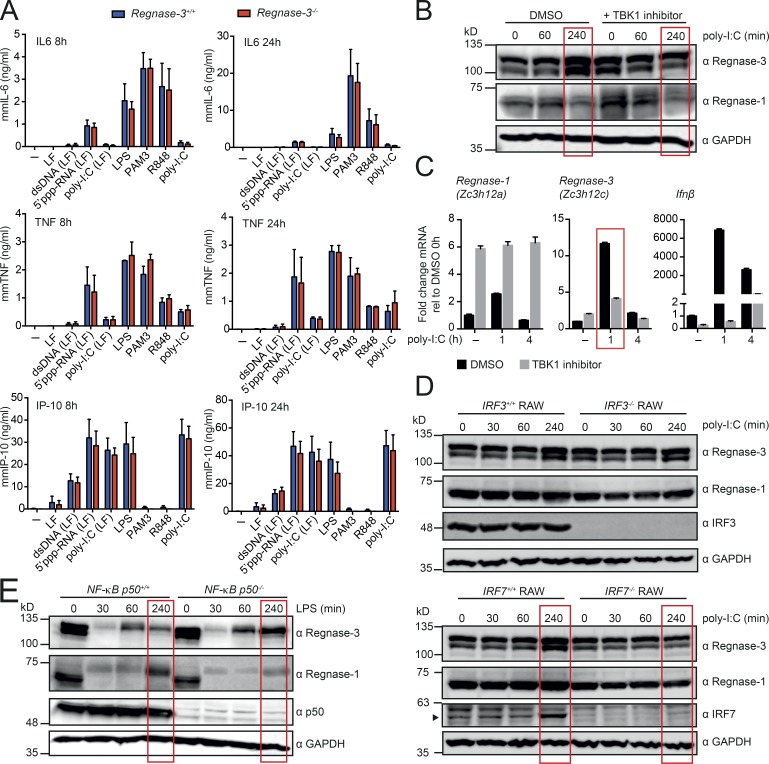
Regnase-3 does not deregulate potential target genes and is regulated via the IFN signaling pathway. (A) BMDMs from Regnase-3−/− mice and Regnase-3+/+ littermate controls were stimulated with dsDNA, 5′ppp-RNA, poly-I:C, LPS, Pam3CSK4 (PAM3), or R848. LF indicates that the compound was complexed to Lipofectamine 2000. 8 h and 24 h after stimulation, ELISA was used to determine IL6, TNFα, and IP-10 in the supernatant. Data are represented as mean ± SEM of three independent experiments. (B) BMDMs from C57BL/6J mice were preincubated for a total of 5 h with Ikkε/TBK1 inhibitor MRT67307 (20 µM) or DMSO (vehicle) and were stimulated with high molecular weight poly-I:C for indicated times and analyzed by immunoblot (representative blot of three independent experiments). (C) BMDMs from C57BL/6J mice were treated as in B, and Regnase-1 (Zc3h12a), Regnase-3 (Zc3h12c), and IFNβ mRNA expression was analyzed by quantitative RT-PCR, normalized to Hprt relative to their expression in untreated BMDMs (mean ± SEM from three biological replicates). (D) IRF3- or IRF7-deficient RAW cells and wild type RAW cells were stimulated with high molecular weight poly-I:C for indicated times and analyzed by immunoblot (representative blot of three independent experiments). (E) BMDMs from NF-κB p50+/+ and NF-κB p50−/− mice were stimulated with LPS (100 ng/ml) for indicated times and analyzed by immunoblot (representative blot from n = 3/3 mice).

