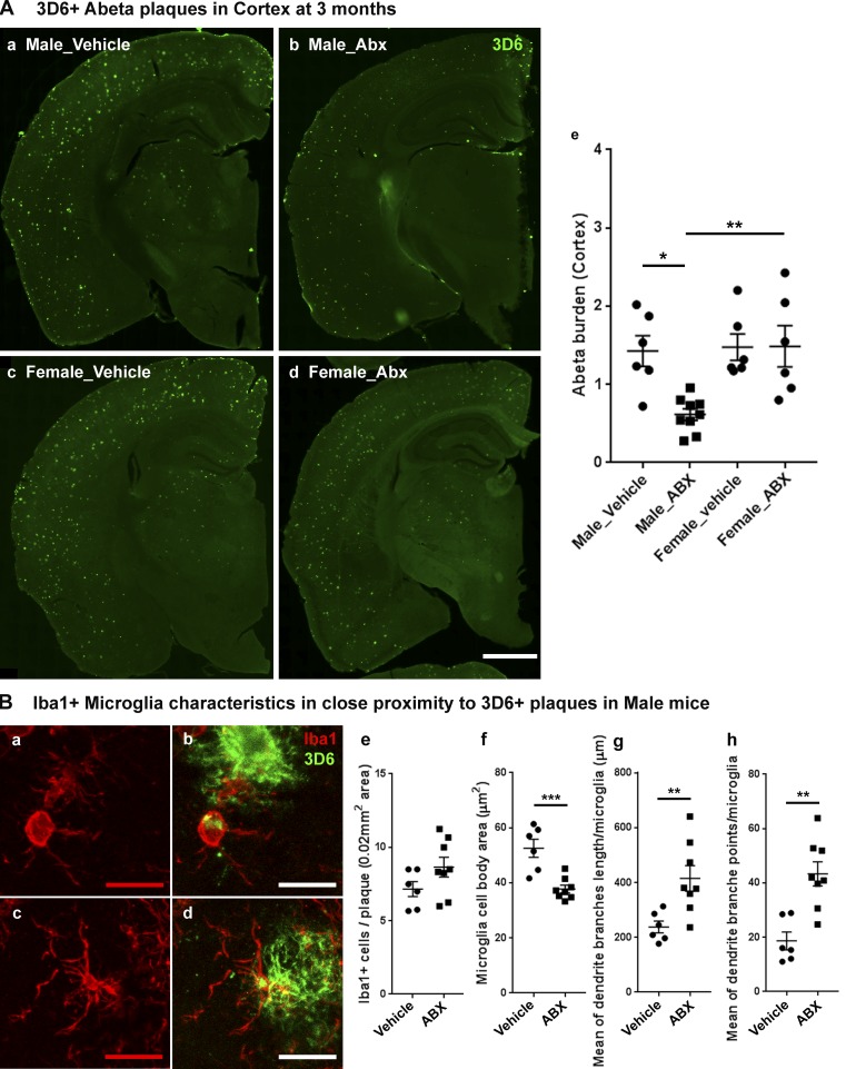
Figure 4.
Reduced Aβ pathology and altered microglial phenotypes in cerebral cortex of ABX-treated male APPPS1-21 mice at 3 mo of age. (A) Representative images of Aβ plaque burden in the vehicle (a and c) and ABX (b and d) treated male (a and b) and female (c and d) mice using 3D6 antibody (e). Aβ burden was significantly lower in ABX-treated male mice (n = 8) compared with vehicle-treated male mice (P = 0.0116) while female mice showed no significant differences in Aβ burden (P > 0.05) with ABX treatment (two-way ANOVA—ABX treatment: F[1, 23] = 5.419, P = 0.0291; sex: F[1, 23] = 7.173, P = 0.0134, and an interaction: F[1, 23] = 5.707, P = 0.0255). (B) Representative images (a–d) of Iba1-positive microglial cells in close proximity to 3D6+ immunostained Aβ plaque (<0.07-mm radial distance from plaque) in 3-mo-old male mice. (e) Cell count analysis showed no significant differences between vehicle- or ABX-treated male mice; (unpaired Student’s t test: t[12] = 1.661, P = 0.1225). ABX treatment (c and d) resulted in smaller cell bodies compared with vehicle-treated (a and b) male mice (f; unpaired Student’s t test: t[12] = 4.573, P = 0.0006). Imaris 3D reconstruction (g and h) showed significantly different microglial characteristics between vehicle- and ABX-treated male APPPS1 mice. Branch length/microglia (g) showed significantly higher branch length in ABX-treated mice (unpaired Student’s t test: t[12] = 3.096, P < 0.0093). Moreover, ABX-treated mice also showed significantly higher numbers of branch points per plaque localized microglia (h; unpaired Student’s t test: t[12] = 4.178, P < 0.0013). Scale bars in A (a, b, c, and d) and B (a, b, c, and d) represent 1,000 µm and 10 µm, respectively. Data are mean ± SEM. *, P < 0.05; **, P < 0.01; ***, P < 0.001. Male_vehicle: n = 6. Male_ABX: n = 8. Female_vehicle: n = 6. Female_ABX: n = 6.

