Figure 3.
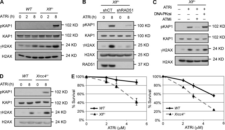
ATR inhibition leads to DDR in Xlf−/− cells. (A) Cell lysates from Xlf−/− MEFs treated with ATRi for the indicated times were analyzed by Western blotting using the indicated antibodies. (B) Cell lysates from Xlf−/− MEFs transduced with Control (shCT) or RAD51 (shRAD51) shRNAs and treated with ATRi for 8 h were analyzed by Western blotting using the indicated antibodies. (C) Cell lysates from Xlf−/− MEFs untreated or pretreated with ATMi or DNA-PKcsi for 30 min before treatment with ATRi for 8 h were analyzed by Western blotting using the indicated antibodies. (D) Western blot analysis of whole cell lysates from WT and Xrcc4−/− MEFs treated with ATRi for indicated times using the indicated antibodies. (E) Cell viability of WT, Xlf−/−, or Xrcc4−/− MEFs treated with the ATRi at indicated concentrations for 4 d. Error bars indicating SD of three technical repeats from a representative experiment from analyses of two independent cell lines of each genotype analyzed in two experiments, each in triplicate.

