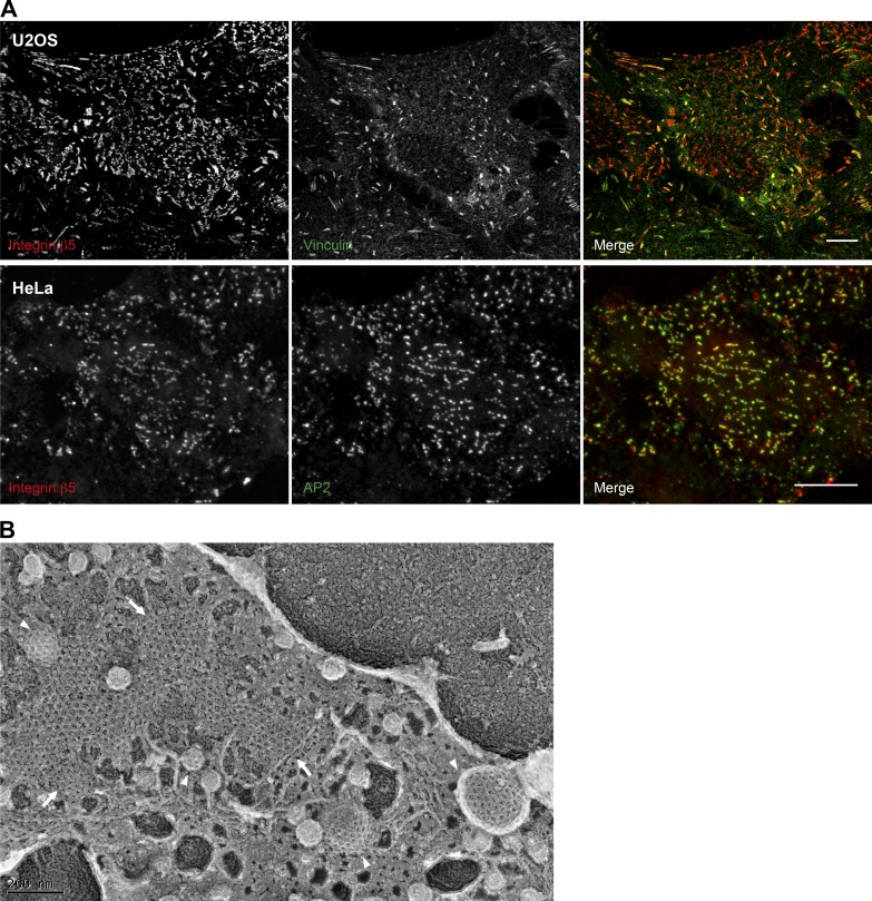
Figure 2.
Imaging of ACs. (A) U2OS and HeLa cells were plated on uncoated or collagen I–coated glass coverslips and cultured for 24 h. Cells were subsequently fixed and immunostained for integrin αVβ5, the canonical AC component vinculin, or the AP2 complex subunit α-adaptin. Scale bars: 10 µm. (B) HS578t cells were plated on collagen I–coated glass coverslips. 24 h later, cells were unroofed by sonication to generate a platinum replica of the inner leaflet of the adherent part of the plasma membrane as previously described (Elkhatib et al., 2017). Imaging was performed by transmission EM. Arrows point to plaques and arrowheads to clathrin-coated pits. Image in B was provided courtesy of Dr. Nadia Elkhatib.

