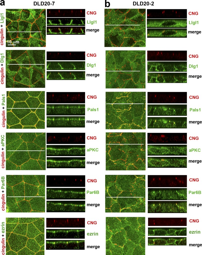
Figure 3.
Polarity proteins in the control DLD20-7 and LAPP-deficient DLD20-2 cells. The confluent 3-d-old cultures of DLD20-7 (a) and DLD20-2 (b) cells were stained with anti-cingulin antibody (CNG, red) and a panel of antibodies against different polarity proteins (green). Among them, two are against Scrib module proteins, Llgl1 and Dlg1, one is against Crb module protein, Pals1, two are against Par6/aPKC complex proteins, Par6B and aPKCζ (aPKC), and one is against the general apical membrane marker, ezrin. A magnification in both the x-y projections and x-z optical sections is the same and is indicated at the top left image. In all images, the central white line indicates position of the Z stacks.

