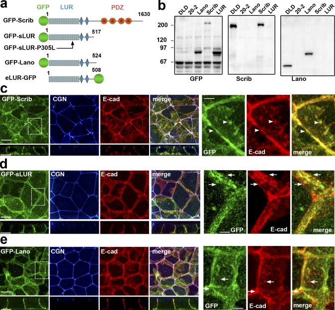
Figure 4.
GFP-tagged hScrib, hScrib-LUR, and Lano are localized at the basolateral membrane and rescue the integrity of AJC. (a) Schematic representation of the recombinant full-length hScrib (GFP-Scrib), its PDZ domain–deficient mutant (GFP-sLUR), its point mutant bearing P305L substitution (GFP-SLUR-P305L), full-length Lano (GFP-Lano), and a PDZ-deficient mutant of Erbin (eLUR-GFP): GFP (green circle); LUR (gray) consisting of 16 LRRs (16 rectangles) and two LAPS domains (diamonds) and PDZ region (red circles). The sizes of the proteins are indicated by the C-terminal amino acid numbers. (b) Western blot of the total cell lysates of the parental DLD1 cells (DLD), the LAPP-deficient DLD20-2 cells (20-2), and DLD20-2 cells expressing GFP-Lano (Lano), GFP-Scrib (Scrib), or GFP-sLUR (LUR). The blots were probed for GFP-tagged proteins using anti-GFP (GFP), hScrib (Scrib), and Lano (Lano). Note that anti-hScrib antibody recognizing C-terminal epitope does not react with GFP-sLUR. Molecular weight markers (in kD) are shown on the left. (c–e) Projections of all x-y optical slices of DLD20-2 cells expressing GFP-Scrib (c), GFP-LUR (d), and GFP-Lano (e). The optical z-sections through approximately the middle of each image are shown at the bottom. Cells were stained for CGN (blue) and E-cad (red) and also imaged for GFP fluorescence (green). Bars, 10 µm. The zoomed areas marked by the dashed line are presented on right panel. Bars, 3.5 µm. Note that majority of AJs associate with GFP-Scrib clusters (arrowheads), but not with GFP-LUR and GFP-Lano (arrows).

