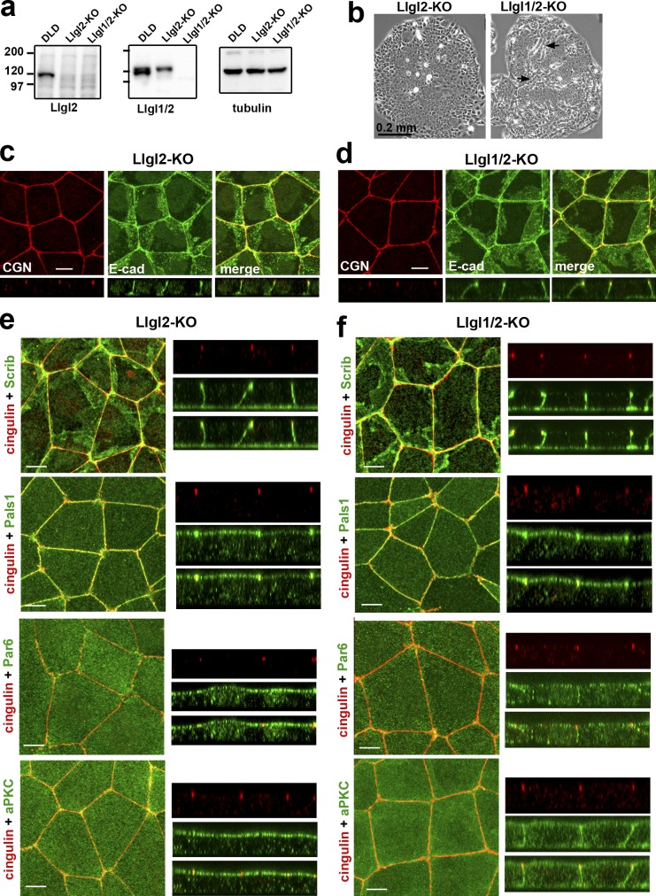
Figure 6.
DLD1 cells deficient for Llgl1/2 exhibit only mild polarity defects. (a) Western blotting of total cell lysates of DLDcas9 cells (DLD), their Llgl2-deficient clone (Llgl2-KO), and the clone of the latter clone obtained after Llgl1 knockout (Llgl1/2-KO). The lysates were probed with antibodies indicated at the bottom. Note that the anti-Lgl1 antibody used in our study (ab18302) cross-reacts with Llgl2. (b) Phase contrast of the Llgl2- and Llgl1/2-deficient colonies (Llgl2-KO and Llgl1/2-KO). Arrows indicate the sites of clear multilayered organization. (c and d) Projections of all x-y optical slices of Llgl2- and Llgl1/2-deficient cells stained for E-cad (green) and CGN (red). The optical z–cross-sections of these images at the same magnification are shown at the bottom. Bars, 10 µm. (e and f) The confluent 3-d-old cultures of Llgl2-KO (e) and Llgl1/2-KO (f) cells were stained with anti-cingulin antibody (cingulin, red) and with antibodies against polarity proteins (green): hScrib (Scrib), Pals1, Par6B (Par6), and aPKCζ (aPKC). Bars, 10 µm. Separate and merged Z stacks for each antibody staining at the same magnification are provided in the right panel.

