Figure 5.
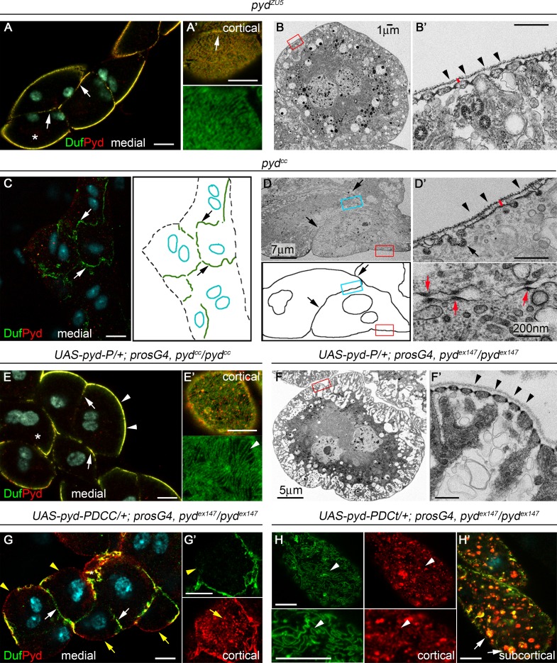
SD formation requires the CC domain of Pyd-P. (A–B′) Confocal (A and A′, n = 150N/10S) and TEM images (B and B′) of larval pydZU5 nephrocytes showing Duf and Pyd at cell junctions (arrows) and normal density of SDs (arrowheads in B′). (C–D′) Confocal (C; n = 147N/12S) and TEM (D and D′) images and schemes of larval pydCC agglutinated nephrocytes. Arrows point to cell contacts, arrowheads in D′ to absence of SDs, red arrows in D′ to junctional complexes, and the red bar to thickening of the BM (compare to B′). (E–F′) Pyd-P rescues the lack of SDs in pydCC (E and E′, n = 85N/7S) and pydex147 (F and F′, n = 72N/6S) mutants (arrowheads). Arrows in E point to Duf and Pyd at junctions between slightly agglutinated nephrocytes. (G–H′) Confocal sections of larval pydex147 nephrocytes expressing Pyd-P lacking the CC (G and G′) or the C-terminal (H and H′) domains. Pyd-PΔCC (n = 86N/9S) fails to rescue SDs formation (yellow arrowheads and arrows mark absence of Duf and presence of Pyd in aggregates at the cortical region). White arrows in G point to Duf at cell junctions. Pyd-PΔCt (n = 73N/6S) promotes SD formation (arrowheads in H) and induces accumulation of Duf/Pyd-containing vesicles (arrows in H′). Asterisks indicate the nephrocytes shown at higher magnification. Unless otherwise indicated, black and white scale bars represent 500 nm and 10 µm, respectively. See also Fig. S1.

