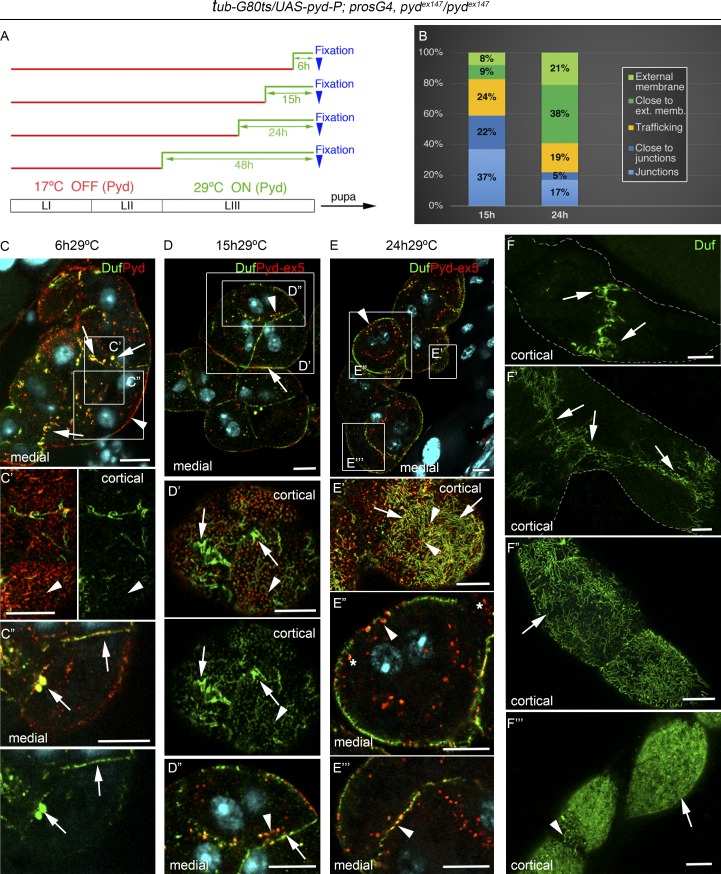
Figure 6.
Pyd-P promotes junctional remodeling and formation of SDs. (A) Chart showing temporal manipulation of pyd-P expression in tub-Gal80ts/UAS-pyd-P; pros-Gal4, pydex147/pydex147 specimens. At 17°C, Gal80ts prevents Gal4-dependent activation of UAS-pyd-P. Nephrocytes were fixed at third-instar larval stage. LI–III refer to larval stages I–III. (B) Quantification of Duf-containing vesicles distribution at 15 h and 24 h of recovery; trafficking class includes vesicles located >2.5 µm away from membranes (n = 16N/3S). (C–E′″) confocal sections of representative nephrocyte strings (C–E) and details of marked regions, of individuals incubated for 6 h (C–C″), 15 h (D–D″), and 24 h (E–E′″) at 29°C before fixation. After 6 h at 29°C (n = 88N/7S), newly synthesized Pyd localizes at discrete puncta in the external membrane (arrowheads in C and C′), and colocalizes with Duf at cell junctions and in large vesicles close to the junctions (arrows in C and C″). After 15 h at 29°C (n = 171N/22S), Pyd colocalizes with Duf at junctions (arrows in D and D″) and in vesicles found mainly close to the junctions (arrowheads in D and D″) and in subcortical regions (arrowheads in D′). Arrows in D′ point to fingerprint-like patterns, indicative of SDs found at external regions close to the junctions. After 24 h of pyd expression (n = 238N/29S), Pyd colocalizes with Duf at SDs all over the cell surface (arrows in E′) at subcortical vesicles (arrowheads in E′ and E″), and occasionally at cell contacts (arrowheads in E″′). Asterisks mark coma-shaped vesicles. (F–F′″) Nephrocyte strings showing progressive recovery of SDs over time (arrows in F″′ correspond to 48 h of recovery; n = 112N/15S). SDs appear first close to the junctions and then extending from there to cover the whole nephrocyte surface. Scale bars represent 10 µm. See also Fig. S4.

