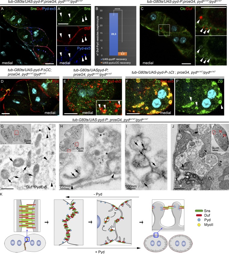
Figure 7.
SD components accumulate in noncoated vesicles near cell junctions and in the external membrane. (A and C–F′) Confocal images of representative pydex147 larval nephrocytes fixed after 15 h (6 h for C) of recovery by expression of UAS-Pyd-P (A, C, and E), UAS-Pyd-PΔCC (D), or UAS-Pyd-PΔCt (F) stained with the indicated antibodies. Vesicles (arrowheads) in A and A′ contain Duf, Sns, and Pyd-P. Duf accumulates in vesicles devoid of clathrin (arrowheads in C; n = 57N/8S). MyoII is detected in puncta in many Pyd-P–containing vesicles (arrowheads in E; the number of MyoII-containing vesicles is significantly reduced by inversion of the green channel, discarding the possibility of random colocalization) and in all Pyd-PΔCt vesicles (F and F′). (B and D) No vesicles are formed after expression of Pyd-PΔCC (n = 63N/7S and 52N/8S for Pyd-P and Pyd-PΔCC). Error bars in B indicate SEM; ****, P < 0.0001 (two-tailed unpaired t test). (G–J) TEM images of pydex147 larval nephrocytes after 15 h of recovery with Pyd-P (G–I) are immunogold stained for Duf and Pyd-ex5. Duf and Pyd-P accumulate at junctions (arrowheads in G), in noncoated irregular-shaped vesicles (arrows in G) larger than clathrin-coated vesicles (asterisks in G, H, and J), at the SDs (arrowheads in H), and in vesicles connected by electron-dense material inside the channels (arrows in I and J). (K) Scheme describing nephrocyte junctional remodeling driven by Pyd-P. In the absence of Pyd, homo- and heterotypic interactions between Duf and Sns might contribute to nephrocyte agglutination. Association of newly synthesized Pyd with Duf at cell contacts favors cis interactions of Duf and Sns in preassembled SD complexes that are sorted into noncoated vesicles. An actomyosin motor is likely involved in trafficking of the vesicles toward the external membrane, where they deliver their cargoes to form SDs. A fall in the levels of Pyd would induce the disassembly of SDs with concomitant changes in Duf–Sns association properties and promote nephrocyte agglutination. Unless otherwise indicated, white and black scale bars represent 10 µm and 500 nm, respectively.

