Figure 1.
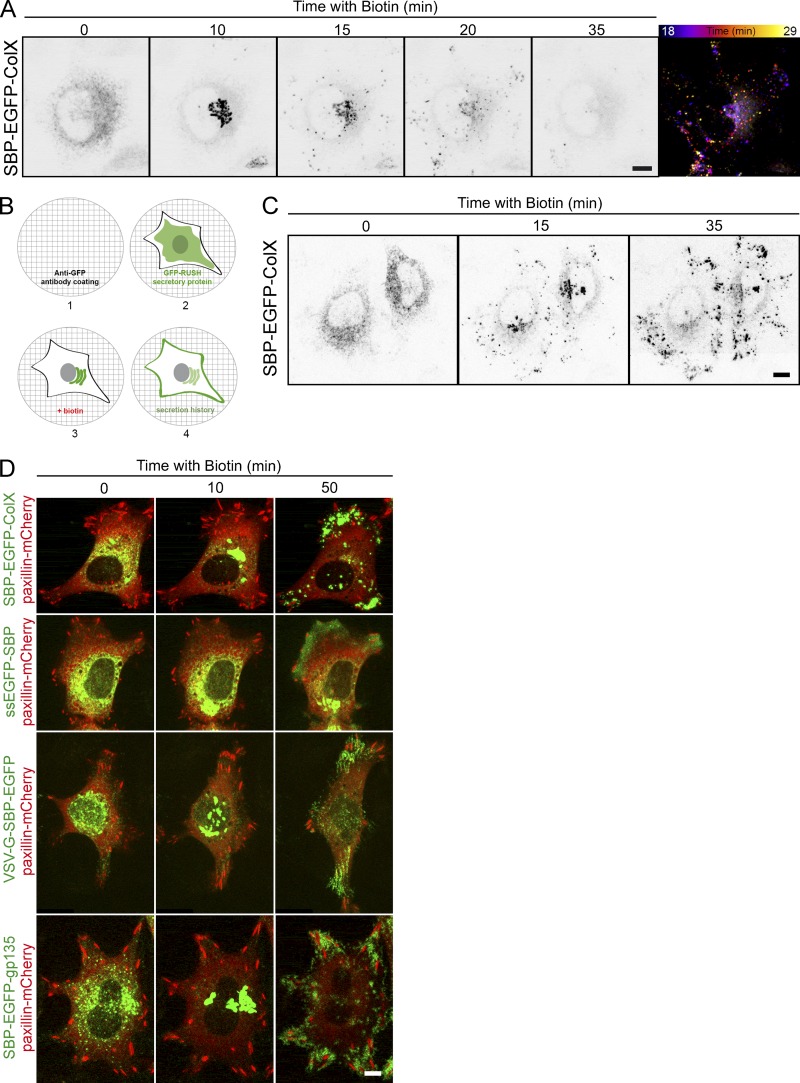
Local exocytosis close to adhesion sites of the cells. (A) HeLa cells stably expressing SBP-EGFP-ColX were incubated with biotin for the indicated time (min). Real-time pictures were acquired using a spinning disk microscope at the indicated times. Temporal projection (right image) was performed for the SBP-EGFP-ColX signal between 18 and 29 min of trafficking. (B) Description of the SPI assay. (1) A coverslip is coated with an anti-GFP antibody. (2) The GFP-RUSH cell line is seeded on the coverslip. (3) Addition of biotin allows the trafficking of the GFP-RUSH cargo. (4) Interactions between the anti-GFP and the GFP of the cargo (transmembrane or secreted) allow the capture of the cargo and provide a picture of the history of the secretion. (C) Trafficking of SBP-EGFP-ColX with an anti-GFP coating (SPI assay). HeLa cells stably expressing SBP-EGFP-ColX were incubated with biotin for the indicated time. Real-time images were acquired using a spinning disk microscope at the indicated times. (D) HeLa cells were transfected with SBP-EGFP-ColX, ssEGFP-SBP, VSV-G-SBP-EGFP, or SBP-EGFP-gp135, and paxillin-mCherry. Coverslips were coated with an anti-GFP coating (SPI assay). Cells were observed by time-lapse imaging using a spinning disk microscope, and pictures were acquired at the indicated times. Scale bars, 10 µm.

