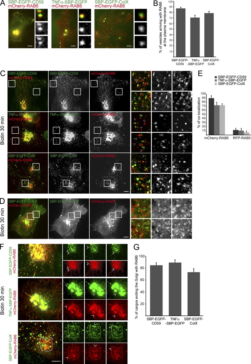
Figure 4.
RAB6 associates with post-Golgi carriers containing CD59, TNFα, or ColX. (A) HeLa cells coexpressing mCherry-RAB6 together with SBP-EGFP-CD59, TNFα-SBP-EGFP, and SBP-EGFP-ColX were incubated for 45 min with biotin and imaged using 3D-TIRF. Representative images taken from the videos are displayed. (B) Quantification of the percentage of RAB6-positive vesicles arriving at the plasma membrane and containing SBP-EGFP-CD59, TNFα-SBP-EGFP, or SBP-EGFP-ColX. Cells were treated as indicated in A (mean ± SEM, n = 217–325 vesicles from 14–18 cells). (C) RPE1-SBP-EGFP-CD59, HeLa-SBP-EGFP-ColX (stably expressing cells or transiently transfected cells), and HeLa-TNFα-SBP-EGFP cells coexpressing mCherry-RAB6 were incubated for 30 min with biotin to allow cargo release from the ER. Cells were imaged using a time-lapse spinning-disk confocal microscope. Representative images taken from videos are displayed. Orange arrowheads point at colocalized vesicles. White arrowheads point at non-colocalized vesicles. (D) HeLa cells coexpressing SBP-EGFP-CD59 and RFP-RAB5A were incubated for 30 min with biotin to allow cargo release from the ER. Cells were imaged using a time-lapse spinning-disk confocal microscope. Representative images taken from videos are displayed. (E) Quantification of the colocalization between mCherry-RAB6 or m-Cherry-RAB5 and each type of cargo (mean ± SEM, n = 9–24 cells). (F) HeLa cells coexpressing mCherry-RAB6 and SBP-EGFP-CD59, TNFα-SBP-EGFP, or SBP-EGFP-ColX were incubated for 15–20 min with biotin and imaged using time-lapse video-microscopy. Representative images of vesicles positive for SBP-EGFP-CD59, TNFα-SBP-EGFP, or SBP-EGFP-ColX and mCherry-RAB6 exiting the Golgi complex together are displayed. Higher magnification of the images taken from time-lapse videos are on the right. (G) Quantification of the percentage of EGFP-SBP-CD59–positive vesicles exiting the Golgi complex with RAB6 (mean ± SEM, n = 13–18 cells). Scale bars, 10 µm.

