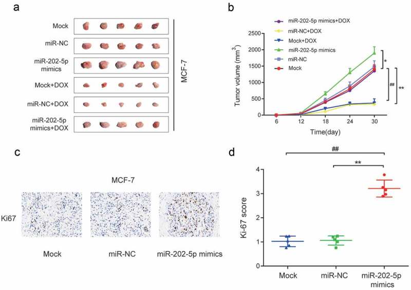
Figure 4.

Overexpressed miR-202-5p promoted proliferation of MCF-7 cells in vivo. (a) Tumor images in different transfection groups. (b) Tumor volume of different transfection groups for 30 days. *P < 0.05, **P < 0.01, compared with miR-NC group. ##P < 0.01, compared with Mock group. . (c) HE stain was performed to detect Ki67 expression in tumor tissues. (d) The graph of Ki-67 scores in different groups. **P < 0.01, compared with miR-NC group. ##P < 0.01, compared with Mock group.
