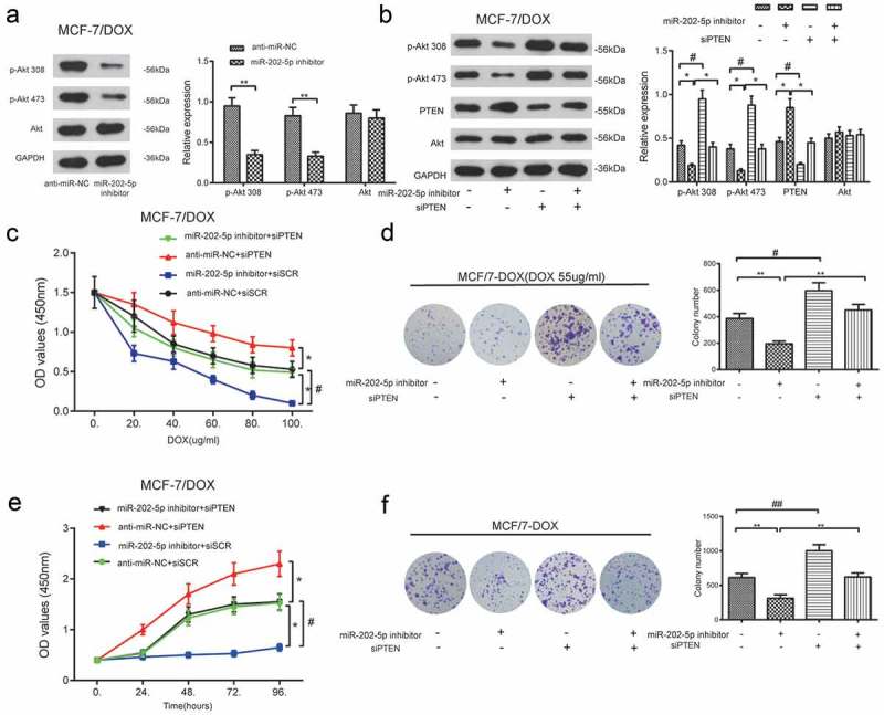
Figure 8.

The inhibition of miR-202-5p decreased MCF-7/DOX cell proliferation through PTEN/PI3K/Akt signaling. (a) The protein expression levels of p-Akt 308 and p-Akt 473 was examined by western blot. **P < 0.01, compared with anti-miR-NC group. (b) The result of western blot showed that the protein expression levels of p-Akt 308 and p-Akt 473. *P < 0.05, compared with anti-miR-202-5p inhibitor group. #P < 0.05, compared with siPTEN group. (c) The OD value of MCF-7/DOX cells was tested by CCK8 assay. *P < 0.05, compared with anti-miR-NC + siSCR group. #P < 0.05, miR-202-5p inhibitor + siPTEN group. (d) The colony formation of transfected MCF-7/DOX cells under the treatment of DOX was shown. **P < 0.01, compared with miR-202-5p inhibitor group. #P < 0.05, compared with siPTEN group. (e) The OD values of transfected cells were measured at 0, 24, 48, 72, 96 h. *P < 0.05, compared with anti-miR-NC + siSCR group. # P < 0.05, compared with miR-202-5p inhibitor + siPTEN group. (f) The colony formation of transfected MCF-7/DOX cells without treatment of DOX was revealed. **P < 0.01, compared with miR-202-5p inhibitor group. ##P < 0.01, compared with siPTEN group.
