. 2019 May 30;8:e46551. doi: 10.7554/eLife.46551

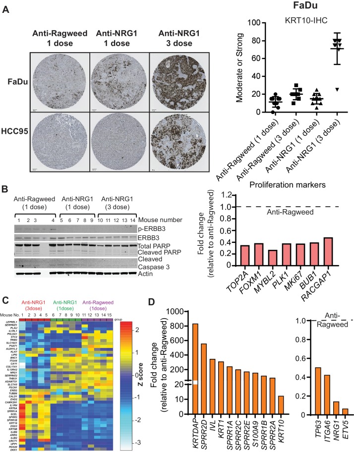
Figure 4. Anti-NRG1 induces squamous differentiation and inhibits proliferation in SCCs.

(A) Representative image of KRT10 (differentiation marker) staining of tumor from FaDu and HCC95 SCC models upon anti-NRG1 treatments compared to anti-Ragweed control. N = 5 mice/group. (B) Protein levels of apoptosis markers upon anti-NRG1 treatment in vivo in HCC95 SCC xenografts. phospho-ERBB3 level was used to assess inhibition of signaling. Expression of proliferation markers upon one and three doses of anti-NRG1 relative to anti-Ragweed treatment in vivo in HCC95 lung SCC by RNAseq. (C) Expression of lung basal cell differentiation markers after one or three doses of anti-NRG1 and one dose of anti-Ragweed treatment in HCC95 lung SCC xenograft tumors by RNAseq. N = 5 mice/group. Expression of (D) squamous differentiation markers and progenitor cell related markers following three doses of anti-NRG1 relative to anti-Ragweed treatment in HCC95 lung SCC xenograft tumors by RNAseq. Average fold change relative to anti-ragweed from n = 5 mice/group.

Figure 4—source data 1. Panel of human airway basal cell gene signature in Figure 4C (as published by Hackett et al., 2011).
DOI: 10.7554/eLife.46551.011

Figure 4.

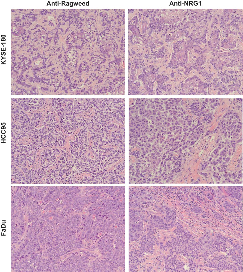
Figure 4—figure supplement 1. Anti-NRG1 induces differentiation in SCC.

Figure 4—figure supplement 1.

Representative image of H and E staining of KYSE-180, HCC95 and FaDu tumors upon anti-NRG1 treatments compared to anti-Ragweed control. N = 5 mice/group.
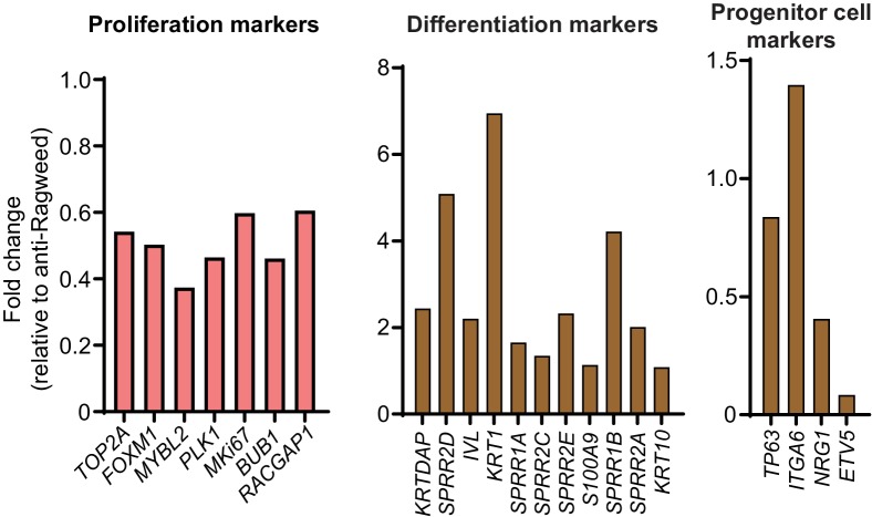
Figure 4—figure supplement 2. Anti-NRG1 inhibits proliferation and induces differentiation in SCC.

Figure 4—figure supplement 2.

Expression of proliferation, differentiation and progenitor cell related markers following one dose of anti-NRG1 relative to anti-Ragweed treatment in HCC95 lung SCC xenograft tumors by RNAseq. Average fold change relative to anti-Ragweed from n = 5 mice/group.
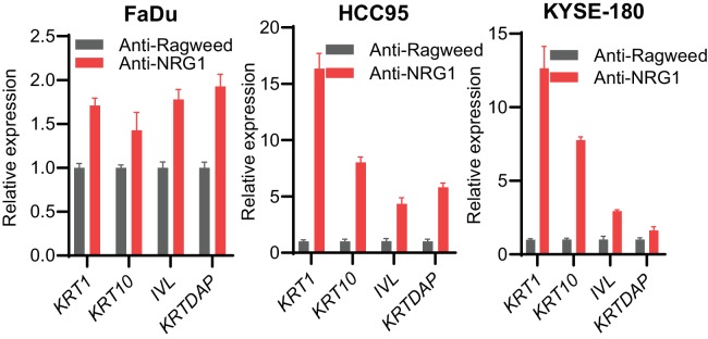
Figure 4—figure supplement 3. Anti-NRG1 increases differentiation markers in vitro in SCC.

Figure 4—figure supplement 3.

Relative expression of differentiation markers such as KRT1, KRT10, IVL and KRTDAP in FaDu, HCC95 and KYSE-180 cell lines after treatment with antibodies for 3 days in vitro.