. 2019 Jun 4;8:e43877. doi: 10.7554/eLife.43877

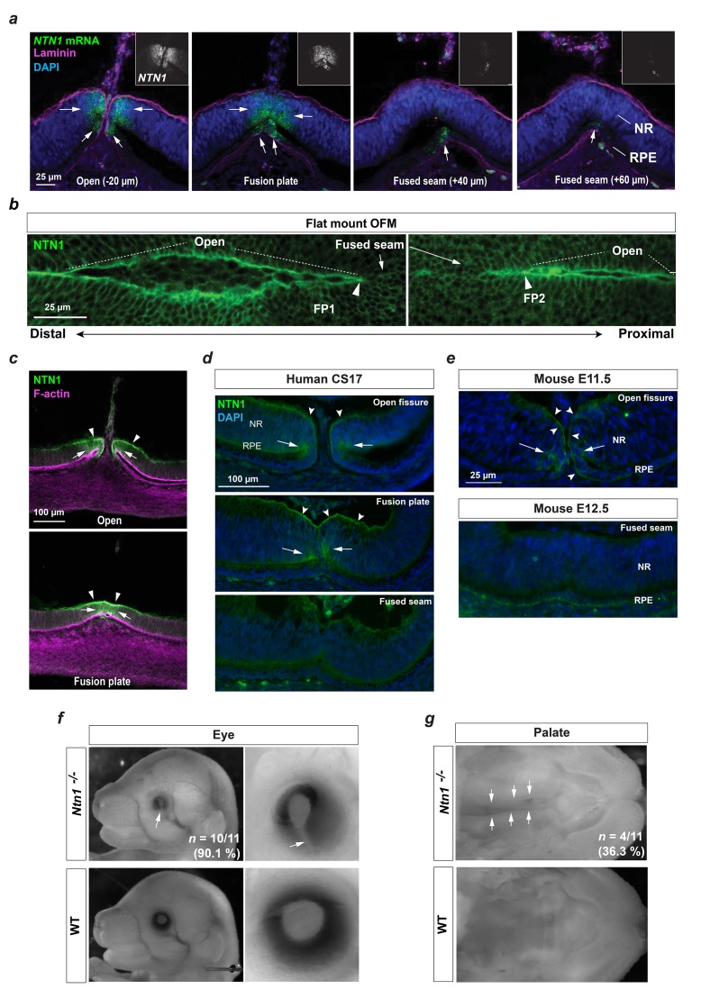
Figure 4. A conserved fusion-specific requirement for NTN1 in OFC and palate development.

(a) RNAscope analysis of NTN1 mRNA (green, and grey in insets) in HH.St29 OFMs revealed fissure-specific NTN1 expression (arrows) with strongest signal observed at open regions and in the fusion plate, and reduced expression in the adjacently fused seam. NTN1 expression was localised to cells of both the NR and RPE. Fusion progression was indicated using anti-laminin co-immunofluorescence (magenta). Images shown are maximum intensity projections of confocal Z-stacks. (b) Single-plane confocal images of immunofluorescence analysis for NTN1 on flat-mounted distal (FP1) and proximal (FP2) OFM revealed enriched protein localisation at the edges of the open fissure margins and reduced in the fused seam. (c) Immunostaining on cryosectioned OFM at the open and fusion plate at HH.St29 revealed NTN1 was specifically localised to the basal lamina (arrowheads) and to the epithelia of the neural retina and RPE (arrows) at the OFM. (d) Immunostaining on CS17 human foetal eye sections revealed human Netrin-1 (hNTN1) was localised to NR epithelia (arrows) and at the overlying basal lamina (dented arrowheads) at the fissure margins. hNTN1 was absent from the fused seam epithelia. (e) Immunostaining for mouse Netrin-1 (mNtn1) in during active fusion stages (E11.5) showed mNtn1 was localised at the open fissure margins (arrow) in the basal lamina and to cells at the NR-RPE junction. mNtn1 was absent from this region in fused OFM seam at E12.5. (f) Ntn1-/- mice exhibited highly penetrant (~90%) bilateral coloboma (arrows; n = 10/11 homozygous E15.5-E16.5 animals analysed). (g) Cleft secondary palate (arrows) was observed in ~36% of Ntn1-/- embryos at E15.5-E16.5 (4/11 homozygous animals).

Figure 4.

Figure 4—figure supplement 1. Developmental NTN1 expression profiling in chick eye and OF.
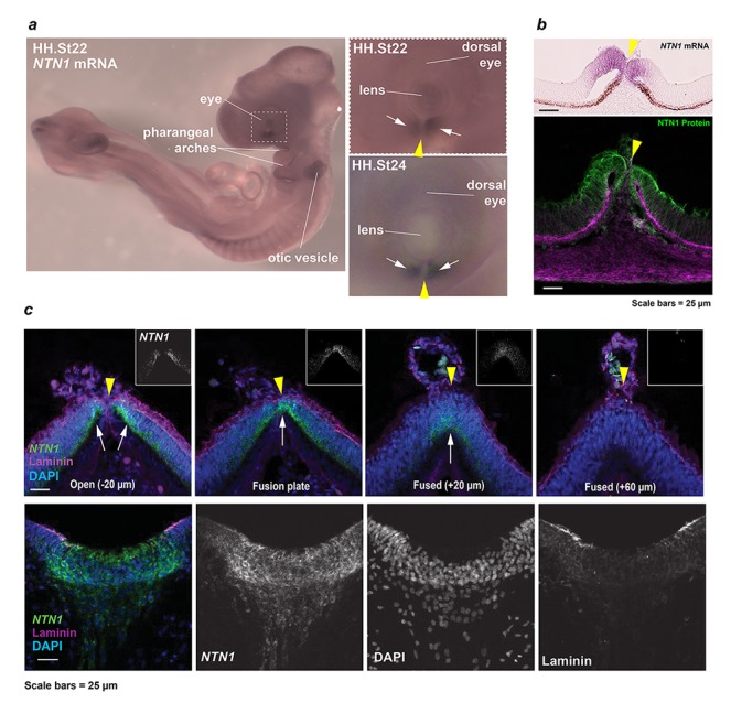
Figure 4—figure supplement 1.

(a) Whole-mount in situ hybridisation revealed NTN1 expression in the ventral eye, developing pharyngeal arches, and otic vesicles at HH.St22. Enlarged panels showed regionally-restricted NTN1 expression in the developing fissure margins (arrows) at HH.St22 (Top) and HH.St24 (Bottom). (b) Section colourimetric in situ hybridisation and NTN1 immunofluorescence analyses at HH.St28 showed NTN1 expression was specific to the edges of the early medial OFM immediately prior to fusion. (c) (Top panels) RNAscope analyses at FP1/distal iris-region OFM at HH.St29 showed NTN1 mRNA specificity (arrows) in the fissure margin and a graded reduction in the fused seam. (Bottom) Positive control analyses for RNAscope showed strong NTN1 mRNA signals in the basal floorplate of the neural tube at HH.St29. The OFM midline is shown by a yellow arrowhead in all panels.
Figure 4—figure supplement 2. Analyses of mouse Ntn1 knockout fissures during fusion.
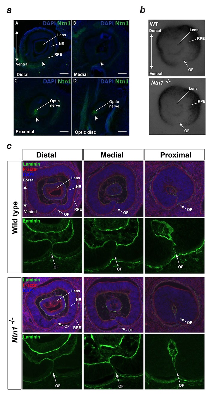
Figure 4—figure supplement 2.

(a) Immunofluorescence staining for mouse Ntn1 in wild-type E12.5 eyes post-fusion showed absence of Ntn1 signal (arrowheads) in the distal (A) and medially fused OFM (B), but presence of Ntn1 (arrowheads) in the proximal (C) and optic disc (D) regions. (b) E11.5 Ntn1-/- embryos did not show any obvious size or gross structural differences during active fusion stages (n = 4 Ntn1-/- embryos analysed, total = 8 eyes). Ventral tissue at the optic fissures (OF, arrows) appeared to be normally apposed. (c) Sections from Wild-type and Ntn1-/- optic fissures immmunostained with anti-laminin antibody (green) and counterstained with DAPI and Phalloidin (red) showed mutant OFMs aligned correctly at E11.5 with no clear structural differences observed between the genotypes. Representative sections from distal, medial, and proximal OFMs are shown. OF, optic fissure; NR, neural retina; RPE, retinal pigmented epithelium.
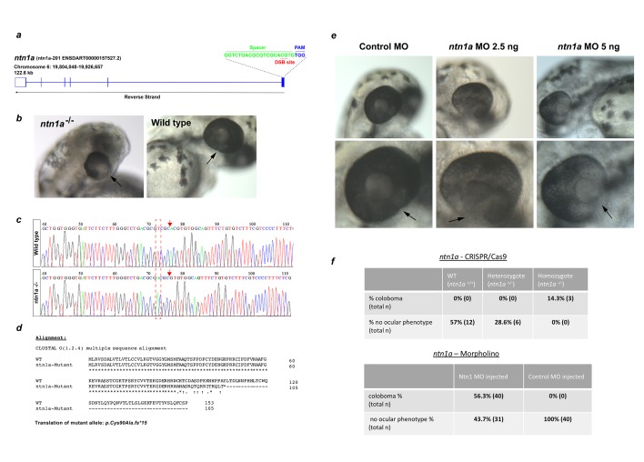
Figure 4—figure supplement 3. Gross ocular phenotype analyses of ntn1-deficient zebrafish.

Figure 4—figure supplement 3.

(a) Gene-editing strategy using a single sgRNA targeting the first exon of zebrafish ntn1a. CRISPR/Cas9 was used to generate heterozygous (ntn1+/-; G0) founders. These were crossed to generate homozygous G1 embryos (ntn1-/-). (b) Panels showing the coloboma microphthalmia and coloboma (arrow) phenotypes in gene-edited ntn1-/- embryos compared to wild-type. (c) Sanger sequencing confirmed the homozygous gene-edited ntn1a allele in 100% of phenotypic G1 embryos. (d) In silico translation of encoded mutant allele aligned to wild-type (first 153 amino acids shown of 603 aa ntn1a protein are shown). The gene edited mutation encodes a frame-shift in the first exon resulting in a truncated ntn1a of 105 amino acids (p.Cys90Ala.fs15). (e) Morpholino experiments produced bilateral coloboma in 100% of embryos injected with ntn1a translation-blocking MO, with no ocular phenotypes observed in control MO injected embryos. The optic fissures are indicated by arrows. (f) Tables with penetrance of colobomas in gene-edited embryos and MO embryos compared to controls.
Figure 4—figure supplement 4. Expression profiling for known interactors of NTN1.
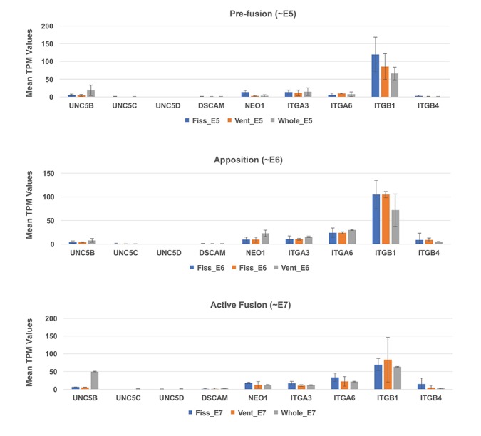
Figure 4—figure supplement 4.

Analysis of TPM values from RNAseq data at all three stages did not detect significant levels of expression for canonical NTN1 receptors in the ventral eye or fissures during OFC stages.
ITGB1 showed the highest expression values throughout all stages, but was not specific to the fissure margin or ventral eye tissues.