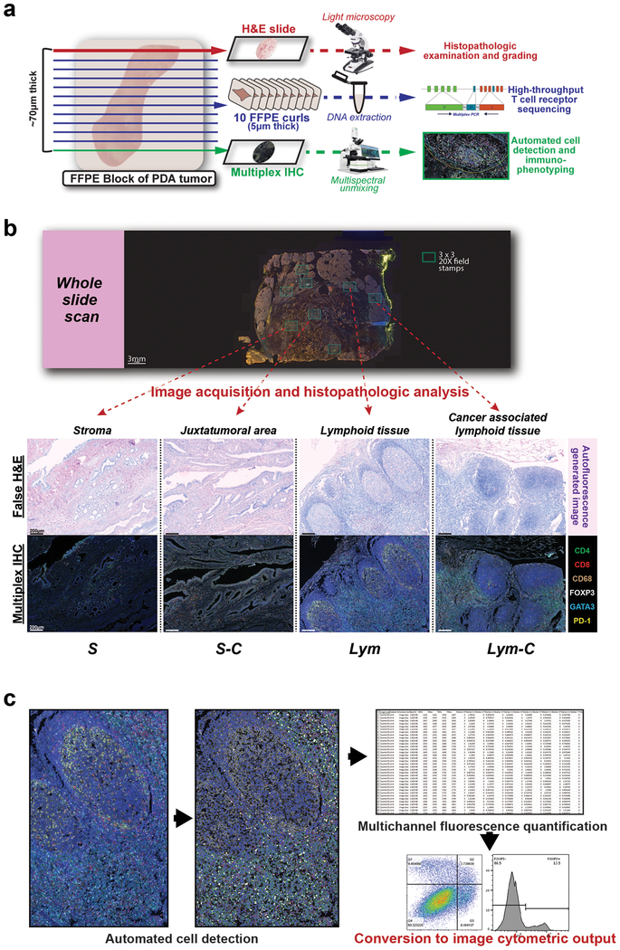
Figure 1: Multimodal histochemical analysis was performed on human PDA resection samples.
(a) Cutting protocol for FFPE blocks of resection specimens of human PDA from patients who were not treated with neoadjuvant therapy (n=30 from 24 patients). Schema shows layout for performing TCR sequencing and mIHC on adjacent curls of the same block. (b) Representative whole slide scan of specimen slide on the Vectra platform were used to evaluate stamps of 3×3 20x high-powered fields (shown in both multicolor IHC and matched autofluorescence-generated false H&E images) to differentiate regions containing carcinoma cells and/or lymphoid tissue. (c) Schema for converting object fluorescence data from multiplex IHC into image cytometric data using FlowJo10.

