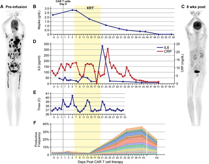
Figure 1. BCMA-targeted CAR T-cell therapy followed by radiation therapy (XRT) led to clinical response, expansion of TCR clonality, and CRS-like findings after XRT.
The patient received conditioning therapy with cyclophosphamide and fludarabine followed by CAR T cells on day 0. Radiation therapy (XRT) took place over 10 fractions between day 6 and day 20 (yellow box). (A) Pre-treatment PET/CT scan showing extensive intra-osseous and extra-osseous FDG-avid disease including soft tissue and pleural-based masses. (B) Decrease in M-spike commencing during XRT. (C) PET/CT 8 weeks post-therapy demonstrating resolution of MM lesions. (D) Prodiuction of IL6 and CRP (pro-inflammatory markers associated with active CAR T-cell function) peaked after the conclusion of XRT. (E) Daily maximum temperature curve revealed a fever at the time of peak IL6 and CRP. (F) TCR clonality analysis demonstrating expansion of novel TCR clones. The subset of TCRs comprising newly expanding clones are shown over time.

