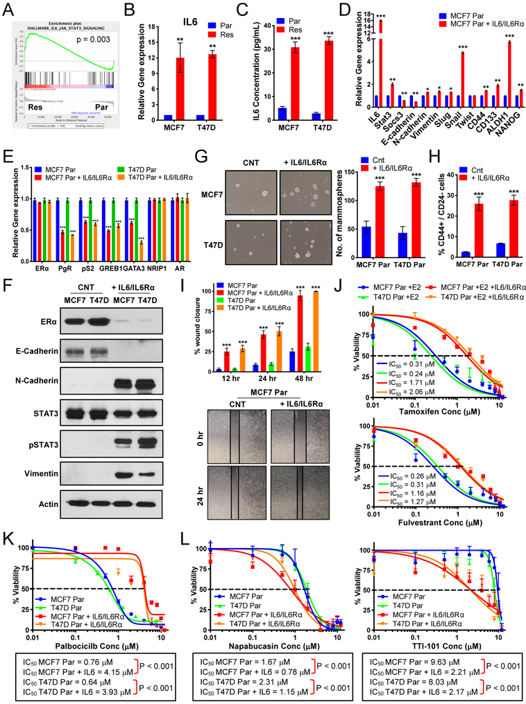
Figure 4: Regulation of the estrogen receptor by IL-6/STAT3 promotes resistance to endocrine therapy and palbociclib.
(A) GSEA enrichment plot of IL-6_JAK_STAT3 signaling indicating increased signaling in resistant cells. (B) qRT-PCR analysis of IL-6 and shows 10-fold increase in MCF-7 and T47D resistant (Res) cells compared to parental (Par) cells. (C) IL-6 ELISA on media collected from MCF-7 and T47D Par and Res cells cultured for 3 days shows a 20-fold increase in MCF-7 and T47D resistant (Res) cells compared to parental (Par) cells. (D) Increased mRNA expression of EMT markers and transcription factors related to breast cancer stem cell-like (B-CSC-L) markers in the parental cells treated with 0.5ng/mL IL-6 and 0.125ng/mL IL-6Rα (i.e. IL-6/IL-6Rα) for days compared to untreated parental (Par) in MCF-7 cell lines. (E) qPCR analysis shows mRNA levels of estrogen responsive genes: pS2, progesterone receptor (PgR), and GREB1, transcription modulators of estrogen receptor: GATA3 & NRIP1, and androgen receptor (AR) in Par treated with IL-6/IL-6Rα in MCF-7 & T47D cell lines. (F) Western blot analysis show levels of ER-α, EMT markers, and STAT3 activation in IL-6/IL-6Rα. treated parental cells. (G) Mammosphere formation is increased in the parental cells treated with IL-6/IL-6Rα. (H) Increased B-CSC-L population observed in the parental cells treated with IL-6/IL-6Rα as identified by CD44+/CD24−. (I) Scratch wound healing assay displays increased cell migration after 12, 24, and 48 hours in the parental cells treated with IL-6/IL-6Rα. (J) Dose response curves in MCF-7 and T47D parental cells (Par) treated IL-6/IL-6Rα after 24hr estrogen deprivation then re-addition of 10nM beta-estradiol (E2) with varying concentrations of tamoxifen (top) or fulvestrant (bottom) (K) Dose response curves show resistance to increasing doses of palbociclib (0.01–12μM) for 6 days and recovery for 6 days in the parental cells treated with IL-6/ IL-6Rα. (L) Dose response curves with TTI-101 (right) and Napabucasin (left) showing increased sensitivity to STAT3 inhibition in parental cells after treatment with recombinant IL-6/IL-6Rα.

