Figure 3.

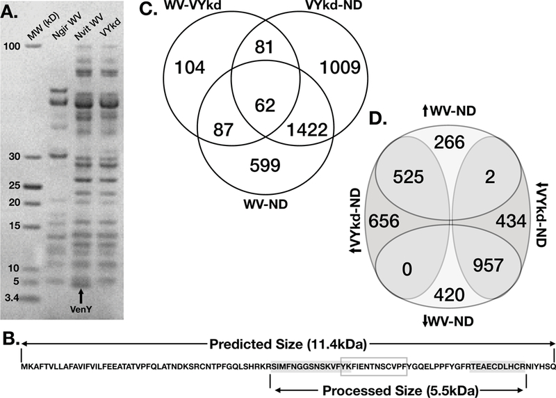
Protein sequencing and knockdown of VenY. a) An SDS PAGE gel shows the size separation of venom reservoir proteins for N. giraulti and N. vitripennis (LacZ RNAi control) compared to N. vitripennis following VenY knocked down via RNAi. VenY (highlighted with an arrow) is the smallest protein visible on the gel, with a molecular weight around 5kDa. b) The amino acid sequence for VenY predicted from the de novo transcriptome assembly of the N. vitripennis venom gland. Peptides sequenced through mass spectrometry are highlighted in grey, which flank the “FIEN” region within the grey box; together they make the predicted 5.55kDa protein. c) Venn diagram of shared significantly differentially expressed genes in the fly host among host injected with whole venom (WV), injected with VenY knockdown venom (VYkd) and unstung normally developing hosts (ND). Venn diagram showing the up- or down- regulation of differentially expressed genes between whole venom and normally developing hosts (WV-ND) and VenY knockdown venom and normally developing hosts (VYkd-ND). The direction of the arrow is in reference to the up- or down- regulation in the first treatment listed.
