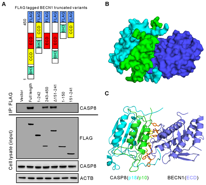
Figure 4.
CASP8 binds to the evolutionarily conserved domain (ECD) of BECN1.
(A) Schematic structures of truncated BECN1 variants used in the experiment (top panel). Immunoprecipitation and western blot of CASP8 binding to FLAG-linked BECN1 variants in DLD1-R cells after transfections with individual plasmids containing BECN1 truncated variants for 48 hours. Cells were lysed and immunoprecipitated with anti-FLAG antibody followed by western blot analysis with the indicated antibodies (bottom panel). (B) Binding model of BECN1 with CASP8. Surface representation of the ECD domain of BECN1 (blue) interacting mainly with subunit p10 (green) of CASP8. The p18 subunit of CASP8 is shown in cyan. (C) Protein-protein interactions at the binding interface. Key residues of BECN1 represented by orange sticks and the p10 subunit of CASP8 represented by yellow sticks are shown in the sticks in brown and yellow, respectively.

