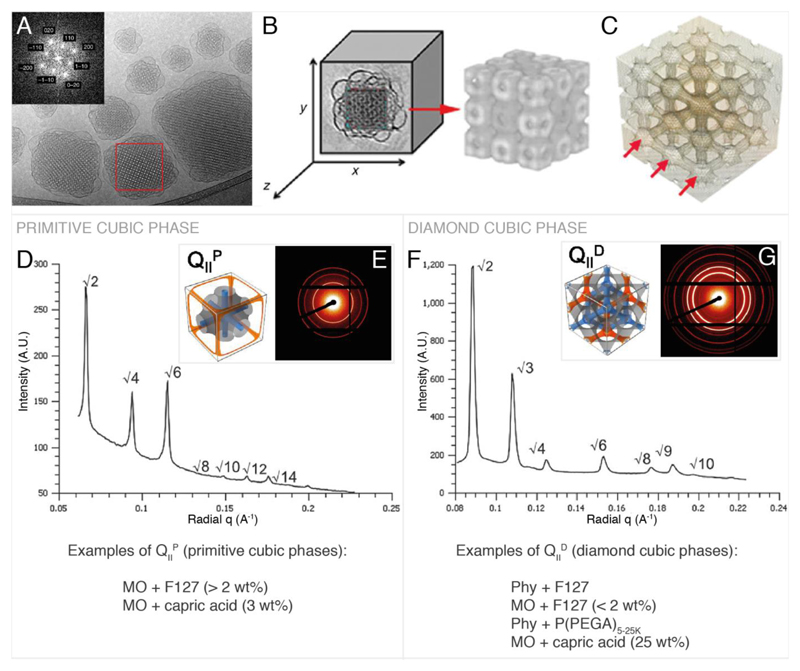
Figure 2.
Primitive and double diamond cubic phases commonly found in cubosomes. A, example Cryo TEM and Fourier transform of cubosomes. B & C, reconstructed 3D image of cubosome from cryo-TEM tomography data showing the lipid arrangement (B) and the water channels (C). D, primitive phase reduced SAXS scattering pattern.[58] E, 2D SAXS scattering pattern[58] corresponding to D and illustration of the primitive cubic phase[45] showing the lipid membrane (grey) and the water channels (blue, orange). F, double diamond cubic phase reduced SAXS scattering pattern.[58] G, 2D SAXS scattering pattern[58] corresponding to F and illustration of the double diamond bicontinuous cubic phase[45] showing the lipid membrane (grey) and the water channels (blue, orange). A, B and C are reproduced with permission from P. Demurtas et al.[33] The reduced SAXS scattering patterns in D & F and the 2D SAXS scattering patterns in E & G are adapted from Ref. [58] with permission from The Royal Society of Chemistry. Illustrations of the primitive and diamond cubic phases in E and G are adapted with permission from H. Kim, C. Leal, ACS Nano 2015, 9, 10214–10226. Copyright (2017) American Chemical Society.

