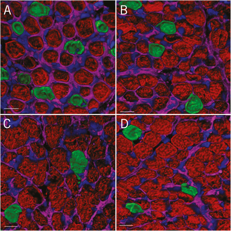
Figure 1.
Confocal laser scanning microscope images of semitendinosus muscle fibers. Cryosections were stained with antibodies to myosin heavy chain (MHC)-fast (red) to identify Type 2 muscle fibers and antibodies to MHC-slow (green) to identify Type 1 muscle fibers. Samples were also counterstained with DAPI to highlight the nucleus (blue) and wheat germ agglutinin (magenta) to decorate the extracellular matrix. Representative images from each treatment group [E−/E− (A), E+/E− (B), E−/E+ (C), and E+/E+ (D)] are shown here.

