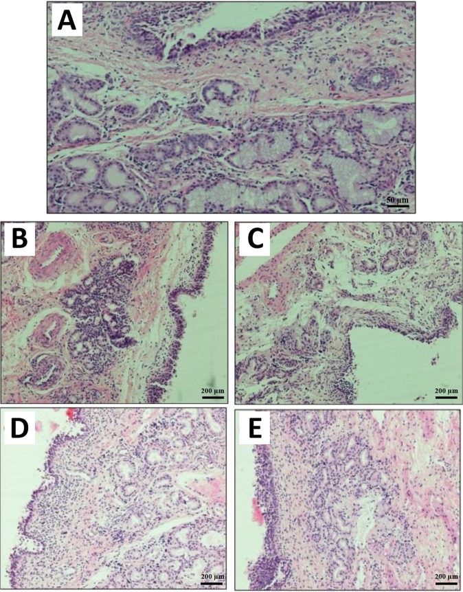
Figure 7.
Histopathological photomicrograph of (A) the normal sheep nasal mucosa (control) (magnification size × 20); (B) nasal mucosa after treatment with hydrogels without additives. Hypotrophy in the Bowman’s gland and cellular infiltration in the connective tissue lamina propria surrounding the gland are perceivable; (C) nasal mucosa after treatment with the PVP + PEG hydrogels; (D) histological characterization of nasal mucosa after using chitosan hydrogel; and (E) nasal mucosa after treatment with thiolated chitosan hydrogel (magnification size × 10).

