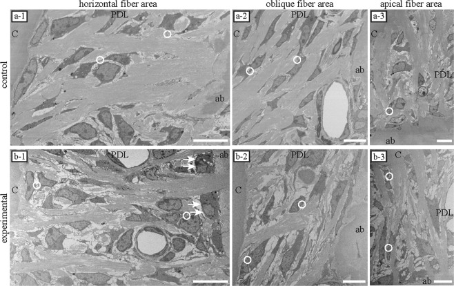
Figure 1.
Single sections imaged via electron microscopy (EM). In both groups, periodontal ligament cells were spindle-shaped and parallelly oriented to the collagen bundle. Cellular processes were observed among the PDL fibres. Cells interacted with adjacent cells (white circle). The area of cellular interaction, which was in contact with other surrounding cells, was narrow. (a-1~3) Collagen bundles tended to be dense in each area of the control group. (b-1~3) Collagen bundles were disorganised in each area of the experimental group. In the area displaying horizontal fibres in the experimental group, osteoblast-like cells were observed on the surface of the alveolar bone (arrows). ab, alveolar bone; PDL, periodontal ligament; c, cementum. Scale bars: 10 μm for all panels.

