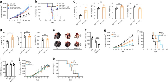
Fig. 6.
Fas signaling relates to the antitumor activity of T helper type 9 (TH9) cells. (a, b) Tumor growth (a) and survival (b) of irradiated WT mice reconstituted with bone marrow cells from wild-type (WT) or Faslpr mice for 2 months and then subcutaneously injected with LLC-OVA tumor cells with or without intravenous injection of 100 µg of anti-interleukin-9 (IL-9)-neutralizing antibodies every other day (n = 5). c Flow cytometric analysis of the frequencies of IL-9+, IFN-γ+, IL-17A+, and Foxp3+ cells among CD4+ T cells in the tumor-infiltrating lymphocytes (TILs) of the mice described in a 20 days after tumor inoculation (n = 3). (d) Enzyme-linked immunosorbent assay (ELISA) measurements of IL-9, IFN-γ, and IL-17A levels secreted by OVA323–339-stimulated TILs from the mice described in a 20 days after tumor inoculation (n = 3). e, f Representative lung appearance (e) and statistical analysis of the lung tumor foci (f) (n = 5) of WT mice 16 days after intravenous injection of B16F10-OVA melanoma cells with no transfer (NT) or transfer of OT-II cTH9 or FasL-TH9 1 and 6 days later. g, h Tumor growth (g) and survival (h) of WT mice that received a subcutaneous injection of B16F10-OVA cells followed by NT or the intravenous injection of 2 × 10⁶ OT-II cTH9 or FasL-TH9 1 and 6 days later (n = 5). i Lung tumor foci of Il9r−/− mice 16 days after intravenous injection of B16F10-OVA melanoma cells with NT or transfer of OT-II cTH9 or FasL-TH9 1 and 6 days later (n = 5). j, k Tumor growth (j) and survival (k) of Il9r−/− mice that received a subcutaneous injection of B16F10-OVA cells followed by NT or intravenous injection of 2 × 10⁶ OT-II cTH9 or FasL-TH9 1 and 6 days later (n = 5). NS, not significant; *P < 0.05, **P < 0.01, and ***P < 0.001 (unpaired Student’s t test: a, c, d, f, g, i, and j; log-rank test: b, h, k). Compared with the Faslpr → WT mice in a, b; compared with cTH9 in g, h, j, and k. Representative results from three independent experiments are shown (mean and s.d.)

