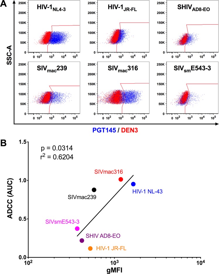
FIG 2.
PGT145 stains cells infected with diverse lentiviral isolates. (A) Overlay plots show PGT145 (blue) versus DEN3 (red) staining of CEM.NKR-CCR5-sLTR-Luc cells infected with the indicated viruses. (B) Area-under-the-curve (AUC) values for ADCC responses were compared to the geometric mean fluorescence intensity (gMFI) of PGT145 staining on the surface of virus-infected cells by Pearson correlation. Virus-infected cells were identified by gating on the Gag+ CD4low population, and PGT145 staining was detected with PE-conjugated anti-human IgG F(ab′)2.

