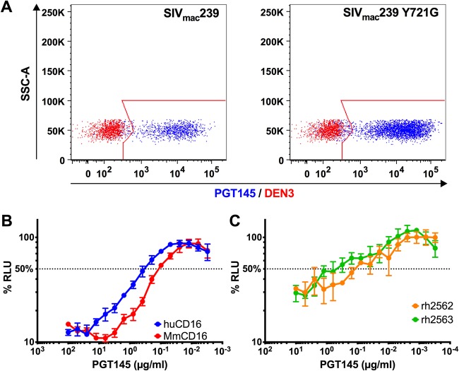
FIG 3.
PGT145 binds to SIV-infected primary rhesus macaque CD4+ T cells and is recognized by rhesus macaque CD16. (A) Overlay plots show PGT145 (blue) versus DEN3 (red) staining of activated rhesus macaque CD4+ T cells infected with wild-type SIVmac239 or SIVmac239 Y721G. Virus-infected cells were identified by gating on the Gag+ CD4low population, and PGT145 staining was detected with PE-conjugated anti-human IgG F(ab′)2. (B and C) ADCC responses were measured using an NK cell line expressing either human or rhesus macaque CD16 (B) or unstimulated PBMCs from two different macaques (C) by incubating SIV-infected CEM.NKR-CCR5-sLTR-Luc cells at a 10:1 effector-to-target-cell ratio in the presence of the indicated concentrations of PGT145. The dotted line indicates half-maximal killing, and the error bars represent standard deviation of the mean from triplicate wells.

