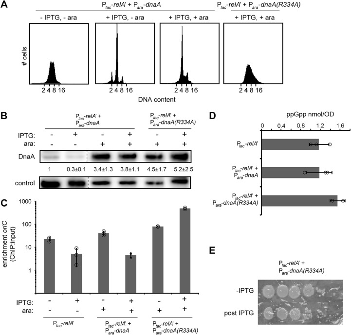
FIG 2.
A decrease in DnaA protein levels is not responsible for the inhibition of replication initiation by ppGpp. (A) Representative flow cytometry analysis of cells harboring Ptac-relA' and Para-dnaA or Para-dnaA(R334A) grown in LB. Overnight cultures were diluted back to an OD600 of 0.01 in fresh media, grown to an OD600 of ∼0.1, treated as follows, and then fixed for analysis: pretreatment (left), RelA′ induction with 100 μM IPTG for 90 min (second from left), DnaA [or DnaA(R334A)] induction with 0.2% arabinose for 30 min followed by RelA′ induction with 100 μM IPTG for 90 min [pDnaA, second from right; pDnaA(R334A), right]. (B) Immunoblots for DnaA in LB for untreated cells and for cells induced for RelA′ and/or DnaA or DnaA(R334A). DnaA levels were normalized to pretreatment levels and quantified from three independent blots. Control is a nonspecific band seen with the DnaA antibody. (C) DnaA association with oriC assayed by ChIP-qPCR enrichment. Relative amount of oriC DNA in a DnaA immunoprecipitate compared to input was quantified by qPCR. RelA′ was induced by addition of 1 mM IPTG for 30 min prior to fixation. DnaA and DnaA(R334A) were induced by addition of 0.2% arabinose for 30 min. oriC DNA levels were quantified using primers against oriC and normalized to a locus near the terminus (relB) that DnaA does not bind. Enrichment was calculated as the ratio of normalized oriC in the ChIP to the input DNA. Error bars represent standard deviation of enrichment from three replicates. (D) Quantification of ppGpp levels in wild-type cells harboring pRelA′, pDnaA and pRelA′, or pDnaA(R334A) and pRelA′. Cells were grown in M9-glycerol plus Casamino Acids, and RelA′ was induced with 100 μM IPTG for 30 min. For pDnaA- and pDnaA(R334A)-containing cells, 0.2% arabinose was added for 30 min to induce dnaA expression prior to addition of IPTG. Error bars are the standard deviation from three replicates. (E) Representative serial dilution plating of E. coli strains carrying pRelA′ and pDnaA(R334A). Cells were grown to an OD600 of ∼0.1, and a sample was taken for serial dilution (top row). DnaA(R334A) expression was induced with 0.2% arabinose for 30 min followed by RelA′ induction with 100 μM IPTG for 90 min, and a second sample was taken for plating (bottom row).

