FIG 5.
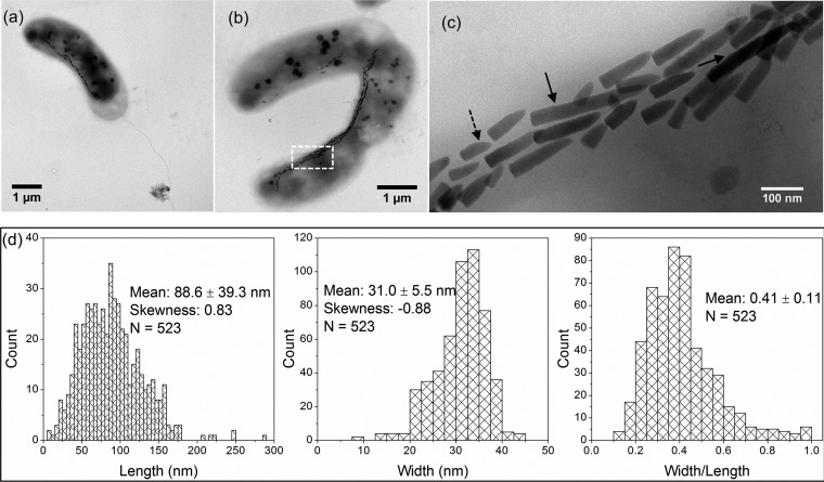
Morphological features of WYHR-1 cells and their magnetosomes. (a) Bright-field TEM image of a bean-like WYHR-1 cell with a rod and a single polar flagellum. (b) Bright-field TEM image of two WYHR-1 cells with intracellular magnetosome chains and spherical phosphate inclusions. (c) Close-up of WYHR-1 magnetosome chains indicated by the white box in panel b. Long (>200 nm) and kinked particles are indicated by solid and dashed arrows, respectively. (d) Histograms of crystal length, width, and width/length ratio of WYHR-1 magnetosomes.

