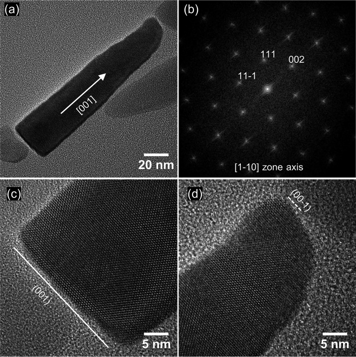
FIG 6.
(a) HRTEM image of a mature WYHR-1 magnetosome from the [1-10] zone axis of a magnetite crystal; the crystal is elongated along the [001] direction. (b) Corresponding indexed FFT (Fast Fourier transform) image for the particle in panel a. (c) HRTEM image of the base of the crystal in panel a, which reveals that the particle is terminated by the (001) face. (d) HRTEM image of the top of the crystal in panel a, which reveals its conical shape.

