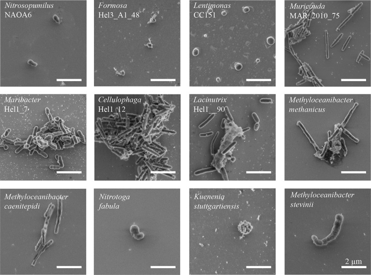
FIG 1.
Representative scanning electron micrographs of the 12 investigated bacterial and archaeal species. All microorganisms were fixed, immobilized on silicon wafers, and critical point dried prior to imaging. The micrographs are ordered according to the cell median volume as follows: Nitrosopumilus NAOA6, Formosa Hel3_A1_48, Lentimonas CC151, Muricauda MAR_2010_75, Maribacter Hel1_7, Cellulophaga Hel1_12, Lacinutrix Hel1_90, Methyloceanibacter methanicus LMG 29429, Methyloceanibacter caenitepidi LMG 28723, Nitrotoga fabula KNB, Kuenenia stuttgartiensis, and Methyloceanibacter stevinii LMG 29431. Scale bars represent 2 μm.

