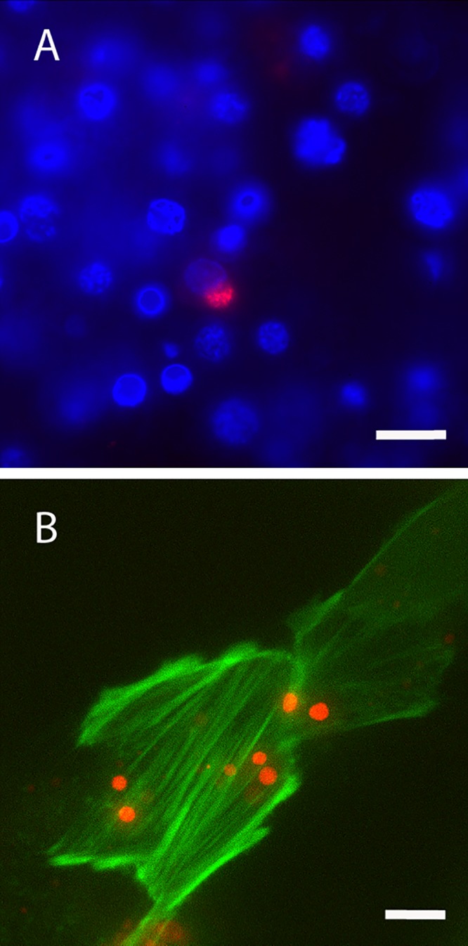
FIG 6.

Live confocal images of mCherry EmCRT in culture. (A) EmCRT infecting ISE6 cells. Ehrlichiae are shown in red, and nuclei are stained blue with DAPI (4′,6′-diamidino-2-phenylindole). (B) EmCRT infecting LifeAct RF/6A cells. Ehrlichiae are red, and host cell actin is green. Scale bars: 20 μm (A) and 10 μm (B).
