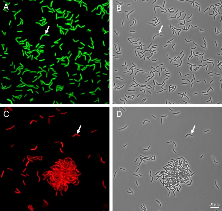
FIG 6.
Fluorescence (A and C) and phase-contrast (B and D) images of the B. cereus ATCC 14579 strain harboring the PcshA′gfp (A, B) and PabrB′mCherry (C, D) fusions. Cells grown at 12°C for 8 h in mAOAC broth were concentrated by a gentle centrifugation and observed with an epifluorescence microscope. Arrows show the same cell in phase-contrast and in fluorescence images. Magnifications, ×1,000.

