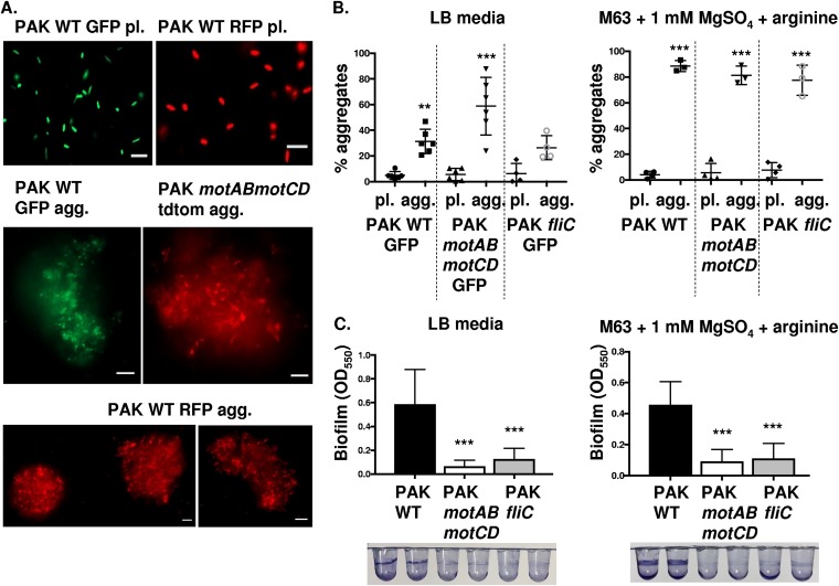
FIG 3.
Wild-type and nonmotile P. aeruginosa (PAK) cells form aggregates, but nonmotile bacteria do not form surface-attached biofilms. (A) P. aeruginosa bacteria (PAK strain) were assayed for the formation of bacterial aggregates. Representative images of PAK WT GFP and RFP planktonic (pl.) bacteria, PAK WT GFP and RFP aggregate (agg.) bacteria, and PAK motAB motCD tdTomato agg. bacteria, as indicated. GFP-expressing bacteria are shown in green, while RFP- and tdTomato-expressing bacteria are shown in red (×20 magnification). Bar, 10 μm. (B) Flow cytometry to assay relative PAK bacterial aggregate formation compared to that of planktonic cultures in LB medium (left) or M63 medium (right). Quantification of aggregate formation is performed by the same methodology as in Fig. 1. (C) PAK WT, motAB motCD, and fliC bacteria were assessed for their ability to form surface-adhered biofilms in the same media as that in panel B. Images of representative wells are shown following staining with 0.1% crystal violet. Biofilm formation was quantified by the OD at 550 nm of the biofilm-associated crystal violet. Data, analyzed using one-way ANOVA with Tukey’s post hoc analysis, are representative of at least three independent biological experiments (n ≥3). ***, P ≤ 0.0005; **, P ≤ 0.005, compared to planktonic bacterial cultures in panel B and compared to PAK WT biofilm formation in panel C.

