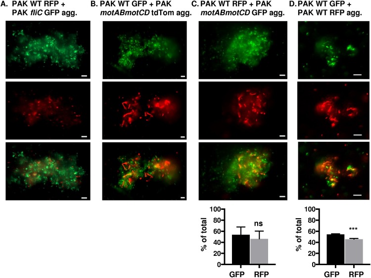
FIG 4.
Wild-type and nonmotile P. aeruginosa (PAK) form coaggregates. Fluorescence microscopy of mixed fluorescently tagged PAK bacteria inoculated together and assessed for coaggregate formation 48 h post mixed liquid inoculation. Representative images of mixed-strain aggregates composed of (A) PAK WT RFP and fliC GFP, (B) PAK WT GFP and motAB motCD tdTomato, (C) PAK WT RFP and motAB motCD GFP, or (D) PAK WT GFP and RFP bacteria, as indicated. GFP-expressing bacteria are shown in green, while RFP- and tdTomato-expressing bacteria are shown in red (×20 magnification). GFP and RFP or tdTomato channels are represented individually in the top and middle rows, respectively, while the combined channels are shown in the bottom row. Bar, 10 μm. (C and D, bottom) Quantification of fluorescent bacteria (GFP or RFP) within aggregates, represented as the percentage of total bacteria. Data were analyzed using an unpaired t test with Welch’s correction (n ≥ 4). ***, P ≤ 0.0005; ns, not significant compared to GFP bacteria.

