FIG 5.
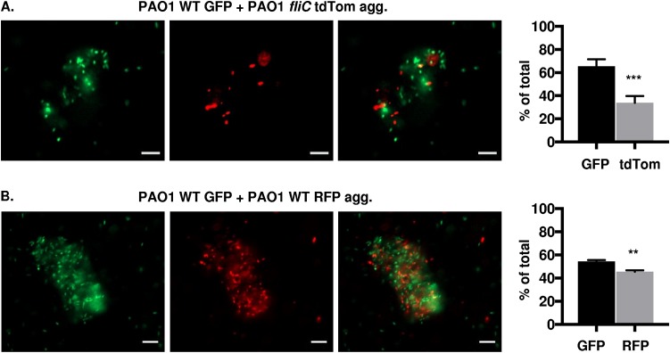
P. aeruginosa (PAO1) cells form coaggregates independent of flagella. Fluorescence microscopy of the indicated strains of PAO1 bacteria, inoculated together and assessed for coaggregate formation 48 h post mixed liquid inoculation. Representative images of mixed-strain aggregates composed of (A) PAO1 WT GFP and fliC tdTomato and (B) PAO1 WT GFP and WT RFP bacteria, as indicated. GFP-expressing bacteria are shown in green, RFP- and tdTomato-expressing bacteria are shown in red (×20 magnification). GFP and RFP or tdTomato channels are represented individually in the first and second columns, respectively, while the combined channels are shown in the third column. Graphs in panels A and B show quantification of fluorescent bacteria (GFP, RFP, or tdTomato) within aggregates, represented as the percentage of total bacteria. Bar, 10 μm. Data were analyzed using unpaired t test with Welch’s correction (n ≥3). ***, P ≤ 0.0005; **, P ≤ 0.005, compared to GFP bacteria.

